What the heck is this? An organism?…an artifact of staining?
A colleague encountered this situation however, with the specimen site in mind and a scan of the smear quickly suspected Streptococcus anginosus group as the culprit in this infection.
Some background;
In 1956, microbiologist W. D. Miller began to isolate and group a number of oral streptococci which later became known as the Streptococcus milleri group, named in his honour. Being streptococci they are gram positive, catalase negative cocci occurring in chains however the milleri group exhibited considerable variability in hemolysis, Lancefield group antigen and other properties typically employed in streptococcal identification. Recently, with the advent of DNA analysis, it was determined that this group consists of three distinct species designated S. anginosus, S. constellatus and S. intermedius. This rearrangement of the S.milleri group is now known collectively as the Streptococcus anginosus group. The historical name of Strepticoccus milleri group, however, continues to remain popular in Britain and parts of Europe.
The Streptococcus anginosus group of organisms are found as normal flora, particularly in the mouth and gastrointestinal tract. As opportunists they have been implicated in systemic infections at multiple body sites and are particularly associated with abscess formation. As such, if isolated from blood culture sterile fluids or abscesses, it is critically important that these organisms not be dismissed as contaminants.
Specifically, alpha or gamma hemolytic members of the S.anginosus group might erroneously be dismissed as the less pathogenic viridians streptococci. Beta-haemolytic members of the S.anginosus group organisms may also be initially confused with S.pyogenes , S.agalactiae, S.equisimilis and large colony group Group ‘G’ streptococci. It is therefore imperative that Streptococcus anginosus group be considered whenever such colonies are evident on cultures of abscesses and normally sterile sites. A complete identification is warranted and the isolates should never be reported solely by their Lancefield grouping. Also, although many of the commercial identification systems, whether manual or automated have the individual Streptococcus anginosus group species in their databases, studies suggest that the accuracy of specific speciation might be as low as 75%. Again, because of the serious pathogenicity of this group, one must be absolutely certain of the identification in reporting.
 Streptococcus anginosus species isolated from Abdominal Fluid (Below)
Clues that an isolate might be one of the S.anginosus group;
- Alpha, beta or gamma streptococci isolated from normally sterile site such as blood*, tissue & body fluids.
- Isolated from abscess.
- Slower growing colonies that may not appear until 36 – 48 hours of incubation.
- Colonies that may exhibit enhanced growth in CO2 or an anaerobic environment.
- Small colonies on sheep blood agar, typically less that 0.5 mm diameter.
- Colonies on agar media give off a characteristic caramel or butterscotch odour.
- Gram stain (see below)
To remember the species comprising the Streptococcus anginosus group, just think of the acronym of America’s Central Intelligence Agency, the CIA where now the letters can stand for Constellatus, Intermedius & Anginosus.
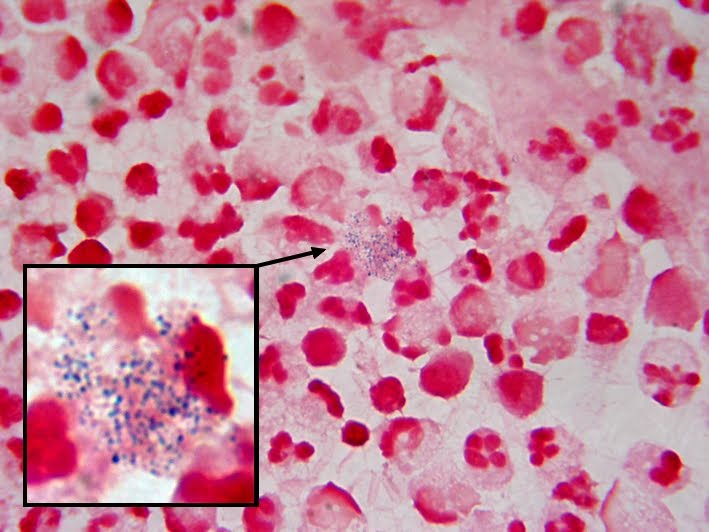 Jumble of Gram Positive Dots within White Blood Cells in Abdominal Fluid
Are these Streptococci?? Do they offer a clue to specific identification?
(Click On Photo To Enlarge for Better Viewing)
 Streptococcus anginosus
Gram Stain of isolated colony taken from Blood Agar Plate of above specimen
This more familiar appearance of Streptococcal cells is what is usually seen even in direct smears of species other than the anginosus group.
 08/01/11: Added this photo of Streptococcus anginosus in a liver asperate (above)
Gram positive cocci in pairs and chains-the textbook description of Streptococcus.
 A quick reference chart of some of the characteristics
the Streptococcus anginosus group may exhibit.
Related Papers;
Streptococcus milleri group: Renewed Interest In An Elusive Pathogen
Streptococcus anginosus (Streptococcus milleri): The Unrecognized Pathogen
* * *

.jpg)


