While I already had a 2010 post on Strongyloides stercoralis , I decided to add a separate posting for this case as it differs in that the worm was not obtained from a fecal sample but rather from a sputum sample. It is precisely these twists that make microbiology both fun and challenging.
An astute colleague was examining a routine sputum culture for bacterial pathogens on this patient when he picked up on a clue that many might have missed. Small looping 'threads' (for lack of a better description) of bacteria which extended from the primary growth (see photo below). He immediately realized that this might be evidence of the presence of a worm. A viable (live) worm if present in the sputum sample would be inoculated onto the agar media along with respiratory bacteria. They can crawl just as an earthworm can crawl on a wet sidewalk. As they migrate around the plate they draw along bacteria that have adhered to their sides and deposit these along the way providing evidence of their travels. With continued incubation the individual deposited bacteria grow up into colonies outlining the worms journey.
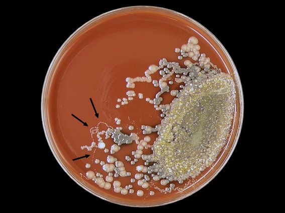 Above (Arrows); A chocolate agar plate inoculated with a sputum sample showing curious looping trails of bacteria which were deposited by a worm migrating about the plate during incubation. Having noticed this curious evidence, the following morning the plate was examined under a plate microscope however the worm responsible for making this trails was not located.
Strongyloides stercoralis can migrate out of the gastrointestinal tract and pass through the lungs. The worm can be coughed out and/or re-swallowed to reinfect the patient. The attending physician had noted that this patient had produced "coffee ground" emesis (hematemesis). This is partially digested blood tinged sputum coughed up, or perhaps swallowed, then coughed up. It has the apperance of wet coffee grounds, hence the description. The sputum sample received by our lab no longer showed this coffee ground appearance. Our patient was an elderly gentleman with several other underlying medical conditions which will not be noted here.

Two Strongyloides stercoralis worms seen in this gram stain of sputum - one curled and one straight directly above. White blood cells dot the background.
Note micron bar in upper right corner. (DMD-108 Microscope X100)
 Same worms as above (Gram Stain X1000)
 Another Strongyloides stercoralis worm (note striated texture of body)
(Gram Stain of Sputum - DMD-108 Microscope: X1000)
 Anterior end of worm with buccal cavity visible.
Compare size to gram negative bacilli bacteria present.
(Gram Stain of Sputum -DMD-108 Microscope X1000)
 Strongyloides stercoralis in Iron Hematolxyin Stained Sputum Isolate.
(DMD-108 Microscope X1000)

Two Strongyloides stercoralis worms seen in this Iron Hematoxylin stained preparation of the sputum sample received. Numerous white blood cells present.
(DMD-108 Microscope X100)
(DMD-108 Microscope X100)

Having requested additional specimen samples (and stool sample for interest sake), Strongyloides stercoralis eggs containing developing larvae were also found.
Above; Both a worm and egg are present with the egg trailing some incidental material attached to one end. (Concentrate of sputum sample DMD-108 Microscope X100)
 Same egg as above with extraneous material trailing off to lower right
(DMD-108 Microscope X400)
 Another Strongyloides stercoralis egg containing developing larvae
(Concentrate - DMD-108 Microscope X400)
 Strongyloides stercoralis worm in sputum.
Above; Both a worm and egg are present with the egg trailing some incidental material attached to one end. (Concentrate of sputum sample DMD-108 Microscope X100)
 Same egg as above with extraneous material trailing off to lower right
(DMD-108 Microscope X400)
 Another Strongyloides stercoralis egg containing developing larvae
(Concentrate - DMD-108 Microscope X400)

(Concentrate - DMD-108 X400)
A stool sample was received by the lab however diagnosis had already been made starting by those rather inconspicuous tracks left by the worm on bacterial agar media.
A stool sample was received by the lab however diagnosis had already been made starting by those rather inconspicuous tracks left by the worm on bacterial agar media.
For further information on clinical symptoms, diagnosis etc, please refer to my previous post located here; Strongyloides stercoralis
Return to top (Most Recent Posts)
.jpg)


