Lots of photos for this one....probably too many, but didn't know what else to do with them!
Hymenolepis nana (Cestode)
–Parasite
Also known as the “dwarf tapeworm”
Geographic
Distribution:
Hymenolepis nana
is a cosmopolitan parasite as it has worldwide distribution.
Associated
Disease:
Hymenolepiasis, or Dwarf Tapeworm Infection. H.nana is often carried by the common
house mouse. While it is more frequently
isolated from children, adults are also quite susceptible. The location of the worm in the infected host
is the small intestine. Light infections
may be asymptomatic however a large worm burden may cause abdominal pain, diarrhoea,
headaches, dizziness and anorexia.
Life Cycle:
Infection usually occurs following the ingestion of H.nana eggs (ova) which make their way
to the small intestine where they subsequently hatch. The released sixed-hooked oncospheres, bury
into the intestinal villi where, after a few days, they develop into
cysticercoid larvae. The cysticercoid
larvae are quite small, containing a single scolex. When mature, the larvae break out of the
intestinal villi to enter the lumen of the small intestine. From the ingestion of eggs to the emergence
of mature worms may take between two to three weeks.
Eggs of H.nana
may also develop into infective cysticercoids in various intermediate hosts,
particularly grain beetles. Accidental
ingestion with contaminated grain products allows the larva to grow into adult
worms in mice and most probably, in humans as well.
Autoinfection is also possible. In this case, eggs passed by the adult
tapeworm hatch within the intestine, develop through the cysticercoid stage and
mature within the intestine as adult tapeworms.
Egg (Ova)
Morphology:
H.nana eggs are
spherical to sub-spherical in shape and have a thin hyaline (clear)
shell. They measure between 30 – 47 µm
in diameter. The six-hooklet oncosphere
is surrounded by a membrane with two polar thickenings, from which arise four
to eight filaments that extend into the space between the embryo and the outer
shell. The related Hymenolepis diminuta
has no polar filaments and this is one feature that aids in their
differentiation.
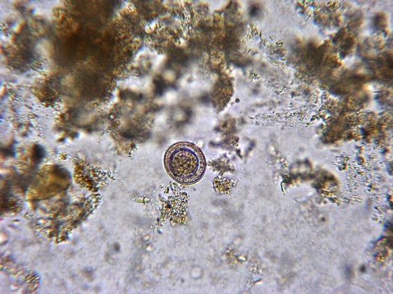
Hymenolepis nana egg (ova): a first look at the fairly low power of 250 times magnification. Here an egg is seen amongst other fecal debris in a concentrated fecal specimen. Care must be taken so as not to over look the parasite as there may be other structures that may mimic or obscure the egg in the concentrate.
(Fecal concentrate, 250X, DMD-108)
Hymenolepis nana egg: A closer view of the H.nana ova in a fecal concentrate.
(400X, Nikon)
Hymenolepis nana egg: Typical appearance of H.nana egg.
(400X, Nikon)
Hymenolepis nana egg: More detail revealed.
(400+10X, DMD-108)
H.nana eggs are
spherical to sub-spherical in shape. They may be described as 'broadly oval'.
Hymenolepis nana egg: Another view - inset showing details.
(400X, Nikon)
Hymenolepis nana egg: Four hooklets are visible in the lower left of the inner oncosphere surrounded by a membrane.
(400X, Nikon)
Hymenolepis nana egg: Another view with the magnified inset showing the position of one of two polar thickenings from which 4 to 8 polar filaments (PF) arise.
(400X, Nikon)
Hymenolepis nana egg: Yet another view.
(500X, Nikon)
Hymenolepis nana egg: Ditto
Hymenolepis nana egg: Now were getting to higher magnifications. Eggs (Ova) measure between 30 – 47 µm
in diameter.
(1000X, Nikon)
Hymenolepis nana egg: Learn to recognize the ova regarless of the orientation and the shape it may be in. (500X, Nikon)
Hymenolepis nana egg: Cell wall appears to be damaged on the left side of this photo. Cell may not be viable, (500X, Nikon)
Hymenolepis nana egg: Oncosphere with a few of the six hooklets are visible in this photo.
(1000X, Nikon)
Hymenolepis nana egg: The pointed structures within the oncosphere (upper part of the inner cellular structure) are the hooklets. Thin, hyaline cellular wall is evident as well.
(1000X, Nikon)
Hymenolepis nana egg: Ditto
(1000X, Nikon)
Hymenolepis nana egg: At least four of the six hooklets are seen within the oncosphere which is surrounded by a membrane. This is contained withing the cell surrounded by the hyaline cell wall.
(1000X, Nikon)
You should be able to recognize the Hymenolipis ova in a fecal concentrate after having viewed all the preceding photos. While the characteristic structures are most clearly viewed in a freshly passed or concentrated fecal specimen, you should be able to recognize the egg in a stained smear as well. A number of photos of iron-hematoxylin stained permanent smears follow.
Hymenolepis nana egg: Here in the same field, one can see that the uptake of the stain and the appearance of the egg can differ significantly.
(500X, Nikon)
Hymenolepis nana egg: At first glance, the egg may even resemble a large amoeba cyst such as Entamoeba coli. Focusing through the cell will not reveal the 8 nuclei expected in E.coli. The size difference also should eliminate the cyst. (1000X, Nikon)
Hymenolepis nana egg: Ditto
(500X, Nikon)
Hymenolepis nana egg: Oncosphere visible, surrounded by a clearing with the outer cell wall barely visible.
(1000X, Nikon)
Hymenolepis nana egg: Oncosphere is visible but cell wall is not.
(1000X, Nikon)
Hymenolepis nana egg: Iron-Hematoxylin stained showing little detail in the oncosphere but cell wall is visible. (1000X, DMD-108)
Hymenolepis nana egg: Another variation of the egg's appearance in an Iron-hematoxylin stained smear.
(1000X, DMD-108)
Hymenolepis nana egg: Dehydration process during staining has distorted the cell wall
(1000+10X, DMD-108)
Hymenolepis nana egg: Egg with outer cell wall and inner ocosphere. Shadows of several hooklets can be seen within, (1000+10X, DMD-108)
Hymenolepis nana egg: Last one.
(1000+10X, DMD-108)
Adult Worm
Morphology:
H.nana adult
tapeworms are quite small, measuring 2.5 – 4.0 cm in length. The tiny, knob-like scolex has four suckers
and a rostellum bearing a ring of 20 - 30 hooklets. Proglottids (segments) are wider than they
are long.
Sorry, I have no worm to take a photo of, however they can be found elsewhere on the web.
Diagnosis:
Diagnosis is made by demonstrating the characteristic
eggs in the fecal sample. Unstained,
concentrated specimens are preferable as the details are more evident. Also, thin-shelled eggs may collapse on
permanent stained smears, making them difficult to identify. Specimens preserved in PVA (polyvinyl alcohol)
do not exhibit morphological characteristic nearly as well as those preserved
in formalin fixed specimens. The adult
worm or proglottid segments are rarely seen in the stool.
Confusion may occur with the related
Hymenolepis diminuta, however
H.dimunata
eggs of the ‘Rat Tapeworm’ are much larger (70 – 85 by 60 - 80 µm in diameter)
than those of
H.nana.
They also do not possess the polar filaments
as previously mentioned.
* * *


.jpg)


