Microscopic
Morphology: M.gypseum has septate hyphae along which sessile or stalked clavate (club shaped) microconidia (3 –
8 µm
X 2 -3 µm)
may be found. The fusiform (spindle shaped) macroconidia (8 – 15 µm
X 22 – 60 µm)
are relatively thin walled, verrucose (with bumpy surface) and contain about 3 –
6 internal cells. M. gypseum’s macroconidia has a rounded apical end while the base
is truncated and may show an annular frill.
They are usually produced in great numbers. This differentiates M.gypseum’s macroconidia from the M.canis macroconidia which has rather pointed ends and the rather
rare & distorted macroconidia produced by M.audounii.Thursday, 17 May 2012
Microsporum gypseum
Microsporum gypseum (Fungus/dermatophyte)
Ecology &
Pathology: Micrsporum gypseum is a
geophilic (soil loving) dermatophyte
which occasionally causes skin infections (tinea
corporis) and/or scalp infections (tinea
capitis). Onychomycosis (nail infections) have also been
reported. It has also been isolated from
horses, dogs, cats and rodents. A cosmopolitan
fungus (found worldwide).
Macroscopic
Morphology: As with other fungi,
colour is influenced by the media it is grown on. Colony generally described as yellowish-buff
to a dark cream or tan colour in colour.
The colony may develop a sterile white ‘feathered’ hyphal border or a
cottony white raised center. The reverse
may be yellow, orange-tan or brownish-red in colour with possible pink to purplish
tinges. Colonies are generally flat with
a granular or powdery texture and exhibit a moderately rapid growth rate,
maturing in about one week.
Microsporum gypseum SAB at 7 Days (Nikon)
Microscopic
Morphology: M.gypseum has septate hyphae along which sessile or stalked clavate (club shaped) microconidia (3 –
8 µm
X 2 -3 µm)
may be found. The fusiform (spindle shaped) macroconidia (8 – 15 µm
X 22 – 60 µm)
are relatively thin walled, verrucose (with bumpy surface) and contain about 3 –
6 internal cells. M. gypseum’s macroconidia has a rounded apical end while the base
is truncated and may show an annular frill.
They are usually produced in great numbers. This differentiates M.gypseum’s macroconidia from the M.canis macroconidia which has rather pointed ends and the rather
rare & distorted macroconidia produced by M.audounii.
Note: Photos that follow were taken with the Leica DMD-108 digital microscope.
M.gypseum -Slide culture at 48 hours (X100 LPCB)
M.gypseum -Slide culture (X250 LPCB)
Hyphae throughout, some showing microconidia, larger macroconidia at left of photo
M.gypseum -microconidia along hyphae are more visible in this photo. Several macroconida throughout. (X250 LPCB)
M.gypseum - another view of both macro & micro conidia (X250 LPCB)
M.gypseum macroconidia (X 400 LPCB)
(Note 100 µm bar at top right of photo)
M.gypseum macroconidia and sessile microconidia attached to hyphae
(X400 LPCB)
M.gypseum clavate microconidia (sessile & stalked?) attached to hyphae
(X1000 LPCB)
M.gypseum - numerous fusiform (spindle) shaped macroconida
(X400 LPCB)
M.gypseum - numerous macroconidia showing up to six internal cells in each. Rough 'verrucose' surface is evident. Adhesive tape preparation. (X1000 LPCB)
M.gypseum macroconidia - on left still attached to conidophore, Rough verrucose surface on lower middle macroconida. (X1000 LPCB)
M.gypseum - more of the same, but I just love them!
(X1000 LPCB)
M.gypsium - macroconidia and a few microconidia (X1000 LPCB)
M.gypseum macroconidium showing rounded apical end and truncated end where once attached to conidiophore. Six internal cells or compartments visible. Rather thin walled with somewhat rough texture evident. (X1000+10 LPCB)
M. gypseum - nearly identical to the macroconidia above, this cell also has six internal cells or compartments with verrucose surface. Nature's own canoe! (X1000+10 LPCB)
* * *
Physiological Tests: M.gypseum
is positive for the hair perforation test.
It requires no special growth factors and produces no change in pH when
inoculated into BCP-milk solids-glucose media.
It is urea positive.
M.gypseum - Intended as computer wallpaper (1024 X 768 when posted)
Aspergillus sydowii
Aspergillus sydowii (Fungus)
Distribution: Worldwide distribution however may occur with
increased frequency between 26o – 35o latitudes. Primarily found in soils but has been
recovered from a variety of sources, both indoor and outdoor.
Pathogenicity:
Implicated as an agent of invasive aspergillosis, keratomycosis (infection of cornea/eye), and
onychomycosis (nail infection).
Aspergillus sydowii SAB 10 Days
Macroscopic
Morphology: Growth rate is moderate.
Colour is influenced by media. It has
been described as a blue-green to dark green to greyish-turquoise. Colonies may have straw-coloured to
reddish-brown shades with exudate.
Reverse is usually maroon, reddish, to reddish-brown. The isolate presented here was grown on SAB media and appears dark green with a white
fringe. Texture has been described as
lanose (woolly), velutinous (dense, silky hairy) or floccose (tufts of hair/hairy). All the preceding means is that A.sydowii produces rather long
conidiophore stipes (~stems) up to
200 µm
which can give the colony that woolly or hairy appearance.
Micsroscopic
Morphology: The long, smooth-walled
stipes which bear the conidiophores are hyaline generally
(translucent/transparent/colourless) or slightly brownish. The vesicles (7.0 µm
- 17 µm wide) may appear sub-spherical, pyriform (pear or teardrop shaped) to
somewhat clavate (club shaped). Conidiogenous structures are biserate with
metulae[1]
(2 µm -3.5 µm by 4 µm -6 µm) and phialides (2 µm – 3 µm by 5 µm – 7 µm) in
size. Diminutive conidial structures are
produced by many isolates which may resemble penicillate (like Penicillium) heads.
Conidia are spherical, echinulate or spinose (rough, jagged texture),
and are about 2.5 µm to 4.0 µm in diameter.
Hülle
cells may be present.
Note: All photos below were taken with the Leica DMD-108 digital microscope. The optical magnification is recorded, however, if a +10 appears after the optical magnification factor, it denotes an additional 10% digital magnification.
Aspergillus sydowii -Slide Culture 48 hrs (X250 LPCB)
A.sydowii - Adhesive Tape Preparation (X1000 LPCB)
Aspergillus sydowii (X1000 LPCB)
Vesicle with biserate conidiogenous structures
Ditto
Aspergillus sydowii - Adhesive Tape Preparation (X1000+10 LPCB)
Vesicle bearing conidiogenous metulae & phailides (biserate)
A.sydowii (X1000 LPCB)
(Note 100 µm bar at upper right)
As above -another view (X1000+10 LPCB)
A.sydowii -Yet another view - As above
A.sydowii (X1000+10 LPCB)
(Note echinulate or spinose [rough] surface texture of conidia)
Aspergillus sydowii (X1000 LPCB)
Aspergillus sydowii - Long Stipes (X1000 LPCB)
A.sydowii - Adhesive Tape Preparation (X1000 LPCB)
(Note reduced conidiophore structure resembling Penicillium species)
A.sydowii conidia (X1000+10 LPCB)
(Note echinulate or spinose [rough] surface texture of conidia)
Note: They may be distinguished from Aspergillus versicolor by their
turquoise colony hue if grown on CYA25 agar.
The moderately growing A.sydowii
produces rather small colonies within a week compared to many other Aspergillus species. The rough conidia borne from small, compact biserate
conidial heads on hyaline to slightly brown stipes is also distinctive.
Sunday, 6 May 2012
Iodamoeba bütschlii
Iodamoeba bütschlii (Intestinal Protozoa – Amoebae) Intestinal Parasite
Distribution: Iodamoeba
bütschlii can be found worldwide however it is less common than Entamoeba coli or Endolimax nana.
Pathogenicity: I.bütschlii
is considered non-pathogenic however it must be correctly identified to
distinguish it from pathogenic intestinal amoebae. Presence of these intestinal amoebae is
evidence that the person carrying it has come in contact with a contaminated
source, raising the possibility of the presence of other parasites.
Trophozoites:
Trophozoites reside in the large intestine where they survive by ingesting
bacteria and yeast but not red blood cells.
In a direct wet preparation made from freshly obtained specimen, the
trophozoites display a sluggishly progressive motility through hyaline
pseudopodia. In an iron hematoxylin or
trichrome stained preparation, trophozoites exhibit a wide range in size,
varying from 6 to 25 µm
with the majority around 9 to 14 µm. Cytoplasm
has a coarsely granular appearance and food vacuoles containing ingested
material may be visible.
The trophozoites have a
single nucleus which is surrounded by a delicate nuclear membrane devoid of
peripheral chromatin. As such, a stained
preparation may not delineate the nuclear membrane and the karyosome may simply
appear to be contained in a vacuole. The
karyosome is large, irregularly rounded and may be central or somewhat
eccentric. With optimally stained preparations, granules
surrounding the karyosome may be visible.
These chromatin granules may arrange to form a ring or radiate outwards between
the karyosome and the nuclear membrane.
Note: Photos of I.bütschlii trophozoites and cysts stained by using the Iron Hematoxylin stain follow. I believe I took all at X1000 magnification. Size may vary between photographs due to my cropping of the photographs. Nikon Coolpix camera used for all but the last photograph.
I.bütschlii trophozoite (Iron Hematoxylin Stain X1000 Nikon)
Ditto
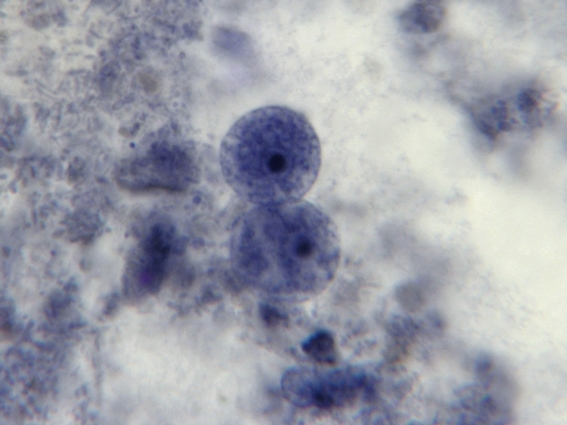
I.bütschlii trophozoite showing large blotchy karyosome withing nucleus. Nucleus outline visible showing no peripheral chromatin.
I.bütschlii trophozoite - cell shape is more irregular that Endolimax nana which is usually rounder.
I.bütschlii trophozoite - again rather irregular shape. Reddish spot (inclusion/surface?) is material which happened to stain acid-fast with the carbol fuschin staining stage added to detect the presence of Cryptosporidium parvum or Cyclospora cayetanesis oocyts if present.
I.bütschlii trophozoite - two cells. Difficult to distinguish from E.nana trophs although generally more irregular in shape and space between nuclear membrane and karyosome appears clearer with fewer chromatin granules.
I.bütschlii trophozoite - Binucleated cell
Cysts: Cysts of I.bütschlii
range in diameter from 6 to 16 µm,
averaging 9 to 10 µm
and may appear spherical however they are usually ovoidal or irregular in
shape. The most striking feature is the
large glycogen vacuole which may contribute to half, if not more of the cysts
size. In fresh specimens stained with
iodine, the glycogen vacuole appears yellow-brown to brown in colour. It is because of this staining property with iodine
that Iodamoeba acquired its
generic name. In an iron hematoxylin
stained preparation, the glycogen vacuole appears clear or off-white’. The cyst nucleus is large, often irregular in
shape, with the karyosome usually eccentric in position. It may even appear pressed against the
nuclear membrane. As with the
trophozoites, the nuclear membrane is devoid of peripheral chromatin and
therefore may not be visible. In well
stained preparations, chromatin granules may be form a crescent shape partially
surrounding the karyosome. Linin fibrils may be seen running between the
karyosome and the chromatin granules.
This arrangement when visible has been described as a ‘basket of flowers’,
with a distorted karyosome forming the ‘basket’, the linin fibrils as the stems
and the granules as the blossoms. Unfortunately my staining and/or the resolution of the camera failed to pick up this fine detail in the photos which follow.
I.bütschlii cyst - Nuclear membrane all but invisible. Large glycogen vacuole seen on right side of cell.
I.bütschlii cyst - large glycogen vacuole takes up most of the cell's interior.
Ditto
I.bütschlii cyst - irregular shaped cell with large glycogen vacuole. Another view.
I.bütschlii cyst - binucleated cell.
I.bütschlii trophozoite & cyst. Cyst nucleus not visible in this view.
I.bütschlii - two cysts.
I.bütschlii -trophs & cyst
I.bütschlii - Cysts in concentrate (unstained)
Trophozoites may be very difficult to recognize in an unstained concentrate as the nucleus is all but invisible. The cyst is more obvious because of its large vacuole which, as above, appears as a void within the cell. Iodine can be added to stain the glycogen, making it more visible with the production of a dark brown colour.
Two I.bütschlii trophozoites seen and one Chilomastix mesnilii trophozoite (just below and to the left of lower I.bütschlii trophozoite) This photo taken with the DMD-108 Microscope/camera.
Diagnosis: Unstained preparations are inadequate for
detecting I.bütschlii as the features
are poorly defined with the nucleus often undetectable. The cyst’s vacuole may be the only structure
visible suggesting the presence of I.bütschlii. Definitive diagnosis can only be made with
the iron hematoxylin or trichrome stained preparation. The cyst form is unique and cannot be mistaken
for any other organism. The trophozoites
however may be mistaken for Endolimax
nana trophozoites as the size range overlaps and both have a similar
nuclear arrangement – a large karyosome (endosome) with no peripheral chromatin
on the nuclear membrane. E.nana trophs are generally smaller, having
a less granular cytoplasm and the space between the karyosome and nuclear
membrane may be devoid of the chromatin granules seen in I.bütschlii.
Differentiation may still be challenging.
A diarrheal sample is more
likely to yield trophozoites rather than cysts.
Epidemiology, Prevention & Treatment: Infective
cysts are transmitted through the ingestion of contaminated food or water. Infection is more likely where sanitary
conditions are lacking. As I.bütschlii is considered to be
non-pathogenic, treatment is not recommended.
Subscribe to:
Posts (Atom)