Saturday, 31 March 2012

Scopulariopsis brevicaulis

Scopulariopsis brevicaulis (Teleomorph: Microascus brevicaulis?*) Fungus

Compare to Scopulariopsis brumptii.

Ecology: Cosmopolitan distribution. The genus has many species - frequently isolated from soil as well as plants and insects.
Pathogenicity: Weakly karatinolytic, S.brevicaulis has been implicated in cases of onychomycosis. Other conditions such as endopthalmitis & endocarditis and skin lesions have also been reported. As with many opportunistic fungi, immunocompromised patients may be at greater risk of infection.
Physiology: The optimal growth range is between 24 – 30oC, with a maximum of 37oC.
Macroscopic Morphology: Colonies (SAB at 30oC pictured below) exhibit moderately rapid growth, reaching maturity in about one week. Colonies are velvety to powdery in texture. Colour may start off as white, quickly becoming a pinkish-brown or buff to cinnamon. Reverse is a honey to golden-brown in colour. Colouration varies with each Scopulariopsis species.
Scopulariopsis brevicaulis on SAB after 5 days at 30oC.
Microscopic Morphology: Hyphae are hyaline and septate. Conidophores extend from hyphae and terminate in brush-like groups of 2 – 4 annellophores (conidiophores). Annelloconidia are globose to ovoid with a truncate base, about 5-9 X 5-7 µm in size and have a fine to coarsely roughened wall texture.
Scopularioposis brevicaulis is frequently encountered in the laboratory and can be distinguished from Scopulariopsis brumptii as the latter has short swollen conidiophores and has a grey to greyish-black appearance macroscopically.
*Teleomorph – sources differ. One states there is no teleomorph for this species while another states Microascus brevicaulis is its teleomorph stage. I’ll leave this discrepancy to be pursued by those interested.
All photos below taken with the Leica DMD-108 Microscope
Note: Size may appear to vary between photos of equal magnification due to cropping of photos prior to posting. The DMD-108 microscope has an additional 10X digital magnification factor that can be added to the optical magnification of each particular objective. While I try to take accurate note of each magnification used, errors on my part will undoubtedly occur when taking perhaps hundreds of photos on each organism. While magnification obviously increases the size of the structure being viewed, you may wish to give more weight to the detail and/or resolution of the the magnification provides.
(Click on any photo to enlarge for better viewing)
Scopulariopsis brevicaulis -Slide culture at 48hrs (LPCB X100)
(not much detail to see but included just for scale)
Scopulariopsis brevicaulis -Slide Culture (LPCB X250)
Scopulariopsis brevicaulis - brush-like structure of annellophores (conidiophore) bearing annelloconidia (LPCB X400)
Scopulariopsis brevicaulis - younger culture showing chains of annelloconidia extending from individual annellophores (conidiophores) (LPCB X400)
Scopoulariopsis brevicaulis - brush-like arrangement of annellophores bearing annelloconidia
(LPCB X400)
Scopulariopsis bdrevicaulis - a closer look at the brush-like structured described above. Note the rough texture on the conidia walls.
(LPCB X1000)
Scopulariopsis brevicaulis (LPCB X1000)
Okay, maybe too many photos, but I always get frustrated while consulting even the best fungal reference books and finding one tiny B&W photo crammed in at the bottom of the page which attempts to illustrate every feature. I think seeing several photos, even of the same structure, gives one a better perspective on the organism's characteristics.
(Ditto)
Scopulariopsis brevicaulis - Brush-like branched annellophores bearing annelloconidia.
(LPCB X1000)
Scopulariopsis brevicaulis Annelloconidia- Okay, last photo. Note the ovoid structure of the annellocondia - 'annello' because of the truncated (flattened) base where they were attached to the annellophore, or simply the conidiophore. (LPCB X1000)

Beauveria species

Beauveria species (probable bassiana) -Fungus

Ecology: A cosmopolitan fungus commonly isolated from soil.

Pathogenicity: Low pathogenicity for humans however reports of keratitis and pulmonary infections have been reported in immunocompromised patients. Commonly considered to be a contaminant. Some Beauveria species are recognized as important insect pathogens particularly as the agent of muscardine disease of the silkworm.

Macroscopic Appearance: Beauveria is a moderately rapid growing fungus. Texture has been described as floccose,velvety, powdery to cottony. The isolate I present below has the appearance of a dollop of shaving cream dispensed from pressurized can. The colour is generally a bone white which can take on a pale yellow tinge with age. The reverse is a non-descript pale colour.

Microscopic Appearance: Hyphae are rather narrow and septate. Conidiogenous cells may grow singly or aggregate in dense clusters along the hyphae. They appear inflated at the base then become thinner at the apex giving a flask-like appearance. At the terminus there is what most sources describe as a thin zigzag filament or extension which bears a conidium at each bend (sympodial geniculate growth). Conidia are single celled, hyaline, smooth walled, round to oval and about 2-4µm in diameter, each singly attached via a fine denticle.

Beauvaria species on SAB after 5 Days at 30oC

(My isolate looked like canned shaving cream)

Beauveria species, slide culture (LPCB X200 Nikon)

Beauveria species, slide culture (LPCB X400 Nikon)

Beauveria species, slide culture (LPCB X400 DMD-108)

(Many clusters of conidiodgenous cells)

Beauveria species, slide culture (LPCB X400 DMD-108)

Beauveria species (LPCB -oops, magnification not noted)

(Resolution at magnification not sufficient to show attachment of conidia)

Beauveria species (oops, magnification not noted)

Beauveria species - both individual conidiogenous cells and clusters seen

I will try to get a higher magnification photo of the conidiogenic cell bearing the conidia from the zigzag like apex. I seem to lose the resolution on the photos or they are so overgrown with conidia that the structure is obscured. Stay tuned...

Home - Return to Most Recent Posts

Saturday, 25 February 2012

Trichophyton rubrum

Trichophyton rubrum (Fungus, Dermatophyte) Teleomporph: None

Ecology, Epidemiology & Pathogenicity;

Trichophyton rubrum is a cosmopolitan, anthropophilic fungus meaning its primary reservoir is man. It is the most widely disseminated dermatophyte of man and most common cause of athlete’s foot (Tinea pedis), jock itch (Tinea cruris), ringworm (Tinea corpis), onychomycosis (Tinea unguium), and less commonly hair and scalp infections. Invasive infections in immunocompromised patients have been reported.

Trichophyton rubrum infections are of concern because nail infections caused by this dermatophyte are extremely difficult to cure.

Presented below is an isolate responsible for a case of onychomycosis, also referred to as Tinea unguium or simply stated a nail infection.

Rate of Growth;

Trichphyton rubrum is a moderately slow grower, reaching maturity within 14 days at 25o to 30oC.

Colonial Morphology;

Trichophyton rubrum species exhibits widely variable colonial morphology and the appearance is further influenced by the media on which it is isolated. Most typical strains are downy to cottony in texture with fine white aerial mycelium at the surface. The overall surface is white, sometimes becoming rose on ageing. The reverse is typically wine-red; however brown & yellow to olive-green hues may be present. SAB & Mycosel media tend to bring out the reddish-brown to yellow colours. Trichphyton rubrum often appears in literature as having two major forms of surface texture; a downy type and a granular type. Overlap between the two may be evident and even taxonomic relatedness is questionable. One source1 states that “deep red isolates with many macroconidia, once referred to as the ‘granular’ colony surface type, are now mostly recognized as representatives of the urease-positive T.raubitshekii.”

Trichophyton rubrum surface and reverse on SAB media after 10 days incubation at 30oC

Microscopic Morphology;

Trichophyton rubrum produces hyaline septate hyphae. The downy type, described in this post, is characterized by the production of moderate numbers of clavate (club shaped) or pyriform (tear-drop shaped) microconidia (3-5.5 X 3-3.5 µm) with rare if any macroconidia. (The granular form is characterized by the production of moderate to abundant numbers of microconida as well as moderate to abundant numbers of long, narrow, thin-walled cigar or pencil-shaped macroconidia (40-55 X 6-7.5 µm) with parallel sides.) Macroconidia (4 – 10 cells in length) may form directly on the ends of thick hyphae singly or in groups. It may also produce small chains of barrel-shaped arthroconida from both the hyphae and macroconidia and are similar in size to the macroconidia.

Photos below were taken with a Digital Nikon Coolpix 8400 Camera or the Leica DMD-108 microscope camera as indicated. The DMD-108 has a digital magnification factor of X10 which can be added to any optical magnification.

Structure size may vary between photographs of identical magnification due to selective cropping of the photograph.

* * *

(Click on any photo to enlarge for better viewing)

Toenail from which this Trichophyton rubrum was isolated. Portions of the nail specimen were placed on SAB and Mycocel media for isolation. Portion was also flooded with 10% KOH which both softens the nail and clarifies for better viewing of any fungal structure which may be responsible for the infection.

Photomicrograph of nail segment in KOH. Trichphyton rubrum fungal hyphae seen invading between nail cells. (dotted structure weaving through from upper left through to lower right) (DMD-108 Microscope X400)

Photomicrograph of T.rubrum fungal element (arrow) in toenail tissue (Magnification not noted)

Trichophyton rubrum microconidia on hyphae. This arrangement of microconida has been described as "birds on a wire" where the pyriform (teardrop shaped) microconidia are attached to the hyphae at the narrow end (tail).

Another view of the microconidia attached to the hyphae (DMD-108 LPCB X400)

Inset: isolated view of the microconidia on hyphae ( LPCB X250 Nikon)

Closer view of pyriform shaped microconidia on hyphae (DMD-108 LPCB X400)

Ditto

This structure looks like I would expect the pencil-like macroconidia would appear, that is except for the size. Though I neglected to take measurements on this structure, it was clearly less than the 45-55 µm usually noted as the average length. Clearly visible are 4 cells within the structure. Perhaps this is still an immature macroconidium.

(LPCB X400 Nikon Camera - I suspect circular lines are from spherical aberrations on the Leica microscope lens at this particular magnification.)

Arrows; indicating presence of Arthroconidia, either free or forming at ends of hyphae,

(LPCB X400 Nikon)

Differentiation;

T.rubrum can be differentiated from T.mentagrophytes by its typical microconidaia which are clavate (club shaped) to pyriform (tear-drop shaped), solitary, sessile alongside undifferentiated hyphae. T.mentagrophytes produces microconidia in clusters that are spherical.

Other Physiological Tests for T.rubrum;

  • Urease Negative
  • Hair perforation test negative.(1)
  • BCP-milk solids-glucose: no change in pH, restricted growth.
  • Potato glucose agar or cornmeal glucose agar enhance the production of red pigment on the reverse if the strain is unpigmented on Sabouraud Glucose (Dextrose) agar.
  • Requires no additional growth factors for growth. Unlike T.megninii, T.rubrum does not require histadine for growth.

Molecular Testing; has been used to reassess the classification of closely related Trichphyton species but remains beyond the scope of the average clinical laboratory and this post.

(1) Hair perforation test; sterile hair can be inoculated with Trichphyton species and examined after a period of growth to see if the fungus has physically invaded/perforated the hair.

Return Home (Most Recent Posts)

Friday, 24 February 2012

Absidia corymbifera (Lichtheimia corymbifera complex)


Mycocladus corymbifer (formerly Absidia corymbifera)
Lichtheimia corybifera complex (formerly Absidia corymbifera) Fungus
(Names seem to change monthly!!! - your choice!) 
Absidia corymbifera;
I’m going to refer to this organism throughout this post by its former name, “Absidia” corymbifera as most are still unfamiliar with the change in nomenclature.
Pathogenicity:
Although uncommon, Absidia species have been implicated as an agent of human (sub)cutaneous mycosis as well as opportunistic pulmonary infections. As with other fungi previously considered to be relatively benign, they are now being isolated with greater frequency particularly in immunocompromised patients (AIDs, transplant patients, neutropenic patients, etc.) Absidia corymbifera is considered to be the only species of Absidia that is recognized as a human pathogen. Absidia corymbifera occasionally has been known to cause mycotic abortions in cows.
Ecology:
Absidia species are ubiquitous in nature and can be isolated from soils and decaying vegetation. As such they may be found as contaminants in cultures. Care must be taken in determining their significance when isolated.
Macroscopic Appearance:
Colonies grow rapidly, maturing in 3 – 4 days. They are white to a greyish-brown in colour with a woolly texture also described as resembling ‘cotton candy’. Reverse is uncoloured and non-descript. Absidia species are inhibited by cycloheximide and therefore will not grow on media such as mycosel. Maximum growth temperature for Absidia corymbifera is between 45 – 52oC. Absidia corymbifera is thermophillic in that it grows more rapidly at 37oC than at 25oC accounting for its human pathogenicity (body temperature). Most other Absidia species fail to grow at 37oC.
Absidia corymbifera on SAB media after 4 days incubation at 30oC
(Reverse not shown - rather nondescript colourless)
Microscopic Morphology:
Absidia’s hyphae are aseptate (rare septa) and relatively wide at 6 – 15 µm in diameter. Absidia species resembles Rhizopus species however the distinguishing feature is that the sporangiophores of Absidia arise from locations on the stolon that lie between the rhizoids and not from them. In other words, there is no rhizoid directly below the sporaniophore as would be observed in Rhizopus species.
Sporangiophores arise alone or in groups from the aerial hyphae and are rather long (up to 450 µm). Sporangiophores branch and widen apically where they form a conical apophysis immediately below the columella. Sporangia are about 20 – 90 µm in diameter are spherical to pyriform in shape. When mature the sporangial wall dissolves releasing round to oval (3 – 5 µm) sporangiospores leaving a small collarette where the apophysis and sporangium had met. Zygospores have been described.
Note: Apophysomyces elegans resembles Absidia corymbifera but produces white colonies and fails to sporulate on routine clinical laboratory media.
Photos below taken with the Leica DMD-108 Microscope.
Young slide culture Absidia corymbifera X100 LPCB
(Click on any photo to enlarge for better viewing)
Absidia corymbifera - Structures seen; (LPCB X100)
  1. Rhizoid
  2. Sporangiophore arises from stolon between rhizoids and not directly from a rhizoid though this appears to be more of a branch in this photo. Rhizoids were a bit of a challenge to find on this particular organism.
  3. Sporangiophore
  4. Sporangium
Ditto (LPCB X100)
Rhizoids were rather difficult to detect. Adhesive tape preparations failed to capture any. I suspect they were so well anchored to the media that the sporangiophores broke off from the stolon rather than pulling the stolon and rhizoids out with it. This photo was taken from a slide culture preparation where some of the agar media surrounding the rhizoid was removed along with the stolon and sporangiophore.
Numerous sporangia with loose sporangiospores are pictured. Notice that the sporangiophore widens apically where it becomes the columella and supports the sporangium. (LPCB X400)
Ditto
Absidia corymbifera sporangium filled with sporangiospores. (LPCB adhesive tape preparation X400 + additional 1% digital magnification)
More views of the same. Apophysis structure can be seen in dissolving sporangium on left. (LPCB X400)
Ditto
Sporangium dissolvingé breaking up releasing Sporangiospores
(LPCB X400)
Apophysis with remains of Collarette and a few remaining Sporangiospores.
More views of same - Stolon with branching sporangiophores bearing Sporangia in various stages of releasing Sporangiospores. Apophysis can be visualized on Sporangiophore at upper left. (LPCB X 400)
Dissolving sporangium releasing numerous sporangiospores with remaining apophysis on apical end of sporangiophore. (LPCB X400)

Wednesday, 22 February 2012

Strongyloides stercoralis Revisited (Sputum)

Parasite (Nematode)

While I already had a 2010 post on Strongyloides stercoralis , I decided to add a separate posting for this case as it differs in that the worm was not obtained from a fecal sample but rather from a sputum sample. It is precisely these twists that make microbiology both fun and challenging.

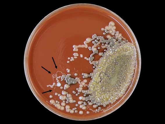
An astute colleague was examining a routine sputum culture for bacterial pathogens on this patient when he picked up on a clue that many might have missed. Small looping 'threads' (for lack of a better description) of bacteria which extended from the primary growth (see photo below). He immediately realized that this might be evidence of the presence of a worm. A viable (live) worm if present in the sputum sample would be inoculated onto the agar media along with respiratory bacteria. They can crawl just as an earthworm can crawl on a wet sidewalk. As they migrate around the plate they draw along bacteria that have adhered to their sides and deposit these along the way providing evidence of their travels. With continued incubation the individual deposited bacteria grow up into colonies outlining the worms journey.

Above (Arrows); A chocolate agar plate inoculated with a sputum sample showing curious looping trails of bacteria which were deposited by a worm migrating about the plate during incubation. Having noticed this curious evidence, the following morning the plate was examined under a plate microscope however the worm responsible for making this trails was not located.

Strongyloides stercoralis can migrate out of the gastrointestinal tract and pass through the lungs. The worm can be coughed out and/or re-swallowed to reinfect the patient. The attending physician had noted that this patient had produced "coffee ground" emesis (hematemesis). This is partially digested blood tinged sputum coughed up, or perhaps swallowed, then coughed up. It has the apperance of wet coffee grounds, hence the description. The sputum sample received by our lab no longer showed this coffee ground appearance. Our patient was an elderly gentleman with several other underlying medical conditions which will not be noted here.

Two Strongyloides stercoralis worms seen in this gram stain of sputum - one curled and one straight directly above. White blood cells dot the background.
Note micron bar in upper right corner. (DMD-108 Microscope X100)

Same worms as above (Gram Stain X1000)

Another Strongyloides stercoralis worm (note striated texture of body)
(Gram Stain of Sputum - DMD-108 Microscope: X1000)

Anterior end of worm with buccal cavity visible.
Compare size to gram negative bacilli bacteria present.
(Gram Stain of Sputum -DMD-108 Microscope X1000)
Strongyloides stercoralis in Iron Hematolxyin Stained Sputum Isolate.
(DMD-108 Microscope X1000)

Two Strongyloides stercoralis worms seen in this Iron Hematoxylin stained preparation of the sputum sample received. Numerous white blood cells present.
(DMD-108 Microscope X100)

Having requested additional specimen samples (and stool sample for interest sake), Strongyloides stercoralis eggs containing developing larvae were also found.
Above; Both a worm and egg are present with the egg trailing some incidental material attached to one end. (Concentrate of sputum sample DMD-108 Microscope X100)

Same egg as above with extraneous material trailing off to lower right
(DMD-108 Microscope X400)

Another Strongyloides stercoralis egg containing developing larvae
(Concentrate - DMD-108 Microscope X400)

Strongyloides stercoralis worm in sputum.
(Concentrate - DMD-108 X400)

A stool sample was received by the lab however diagnosis had already been made starting by those rather inconspicuous tracks left by the worm on bacterial agar media.

For further information on clinical symptoms, diagnosis etc, please refer to my previous post located here; Strongyloides stercoralis
Return to top (Most Recent Posts)










