Monday, 31 March 2014

Hortaea werneckii


Hortaea werneckii -Mould               (Phaeoannellomyces werneckii)

Ecology:
Hortaea werneckii is a saprophytic fungus which can be found in tropical and sub-tropical soils, compost, and decaying wood.  This fungus is also halophilic as it has been isolated from salted foods, saline waters and natural saltpans, growing in salt concentrations of up to 10%.

Pathogenicity:
H.werneckii is the etiological agent of superficial skin infections known as tinea nigra.  This infection usually manifests itself as brown to black macules which are most often found on the palms of the hands.  The appearance may initially resemble that of malignant melanoma.


Macroscopic Morphology:
Hortaea werneckii is a rather slow growing fungus which initially appears as shiny black, slimy or mucoid yeast.  As the colony matures, development of aerial hyphae may give the colony a velvety texture.  The reverse also has a rather non-nondescript brown-black colour.

 Hortaea werneckii SAB 16 days at 30˚C (Nikon)

Reverse: (not shown) appears much like the surface, perhaps slightly more olivaceous in colour.
Right: single colony incubated for approximately one month shows a slight velvety texture.

Microscopic Morphology:
Aerial mycelia develop and acquire the olivaceous black colour as they age.  Septate hyphae may be rather wide, reaching up to 6 µm in width.
Intercalary or lateral conidiogenous (annellide[i]) cells develop along the hyphae which produce the conidia (annelloconidia).  When released the annelloconidia show a prominent annellated ring, 1 -2 µm wide where once attached. 
Annelloconidia are also hyaline (clear) becoming translucent olivaceous brown-black in maturity.  They are smooth-walled and have a broad ellipsoidal appearance (7.0 – 9.5 µm X 3.5 – 4.5 µm).  Annelloconidia are 1 to 2 celled and the internal cell wall or septum is usually deeply pigmented.  With aging the annelloconidia may develop into chlamydoconidia-like aggregates (chlamydospores).

 Hortaea werneckii - At low magnification there is not much to see other that a sea of cells (conidia)
(KOH, 250X, DMD-108)

Hortaea werneckii -brown or olivaceous pigmented conidia are visible
(KOH, 400X,  DMD-108)

Hortaea werneckii - two celled conidia can now be distinguished.
(KOH, 400+10X, DMD-108)

Hortaea werneckii -Pigmented, septate hyphae with lateral annellides.
(LPCB, 1000X, DMD-108)

Hortaea werneckii -Here we can see the broadly ellipsoidal, two-celled annelloconidia.  Mature conidia have the brown pigmentation while younger cells stain more intensely with the Lactophenol Cotton Blue (LPCB).  One end of the annelloconidia usually stains darker indicating the location of the annellated ring or the point where it was previously attached to the conidogenous annellide.
(1000X, LPCB, DMD-108)
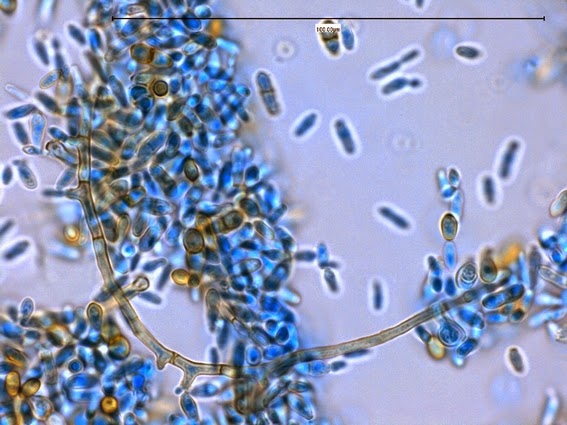
Hortaea werneckii -Another view of a curving septate hyphae surrounded by numerous free conidia.  Annellides can be seen at various points along the hyphae from where the conidia are produced.
(1000X, LPCB, DMD-108)

Hortaea werneckii -The arrow points to an annelide with its developing annelloconidium still attached.  Also seen is a pigmented and septate hyphal element near the top left as well as annelloconidia in various states of maturity.
(1000X, LPCB, DMD-108)

Hortaea werneckii -Another view of a developing annelloconidium attached to the parent annellide.
(1000+10X, LPCB, DMD-108)

Hortaea werneckii -and one more.  Annelloconidium developing in the center of the photo.
(1000+10X, LPCB, DMD-108)
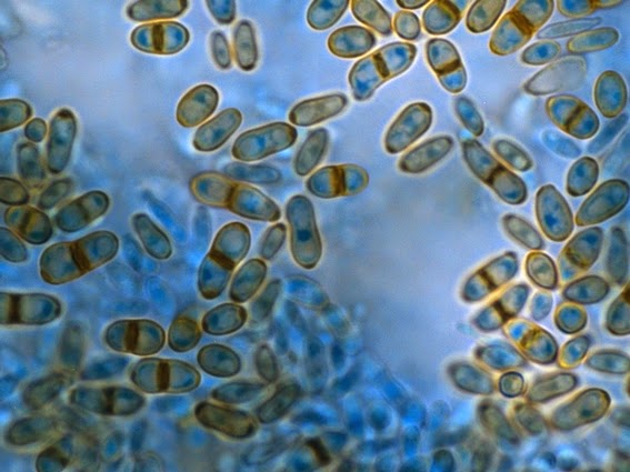
Hortaea werneckii -The primarily two-celled conidia, or more precisely annelloconidia are seen here in various states of pigmentation.  Note that the septum dividing the two cells is darkly stained, as is one end, where the annelloconidium was once attached to its parent annnellide.
(1000+10X, LPCB, DMD-108)

Hortaea werneckii - With aging the annelloconidia may develop into chlamydoconidia-like aggregates (chlamydospores) as seen here in the center of the photograph.
(1000+10X, LPCB, DMD-108)

Hortaea werneckii -The annelloconidia can themselves "bud" and act as annellides, producing annelloconidia as seen here.  
(400X, LPCB, 400X -adhesive tape mount)

Hortaea werneckii -germination of an annelloconidium at top.  Not also the bud developing at the bottom of the two celled annelloconidium.
(1000+10X, LPCB, DMD-108)

Physiology:
-Does not grow at 37˚C
-Nitrate +ve
-Urease +ve
-Halophilic up to 10%

Differentiation:
The annellated zones on Hortaea werneckii are much broader than those of Exophiala species.  Lack of growth at 37˚C also distinguishes the two.


[i] Annellide –A specific conidiogenous cell that produces conidia in succession, each leaving a ring-like collar on the cell wall when released.  ‘Annello’ prefix simply specifies the type of conidiogenous cell or conidia which is produced.

*   *   *

Saturday, 25 January 2014

Vancomycin Dependant Enterococcus (VDE)


Vancomycin Dependant Enterococcus (VDE)

Explanation of Enterococcus faecium’s curious response to the antibiotic Vancomycin

So you try to determine the Minimum Inhibitory Concentration (MIC) of Vancomycin against an Enterococcus isolate by E-test (epsilometer test) methodology and after appropriate incubation you obtain this curious result:

Enterococcus faecium's response to a Vancomycin E-test
(Mueller-Hinton Agar - 24+hours, 37˚C)

What the *$%#&!;.....??  The greatest growth is where the antibiotic concentration is the greatest and tapers off where the antibiotic concentration is the lowest.  It is kind of like shooting at a flock of ducks flying overhead, and only the ones that don’t get hit drop!!!

So what is happening here?  Let’s back up a bit and get some history:

Vancomycin is an important antibiotic as it is the last ‘common’ antibiotic active against most gram positive organisms.  Once an organism acquires resistance to vancomycin, the antimicrobial arsenal is greatly limited in what can be used to fight an infection.

Vancomycin resistance is plasmid mediated, meaning that vancomycin sensitive enterococci and acquire resistance from other organisms already vancomycin resistant.  In turn, these Vancomycin Resistant Enterococci (VRE) can pass the plasmid on to other organisms.  This may result in an outbreak of resistant organisms which are challenging to treat and may be particularly devastating in severely debilitated patients.

There are eight known vancomycin resistance genotypes in enterococci with those known as Van-A being most prevalent, followed by Van-B.  Van-C offers low level intrinsic resistance to E.gallinarum & E.casseliflavus.  The remaining genotypes have not proven to be significant in the clinical setting.

Enterococci expressing the Van-A genotype are resistant to both vancomycin & teicoplanin.  Expression of the Van-B genotype conveys resistance to vancomycin but the enterococcus remains susceptible to teicoplanin.  Van-A resistance is generally higher (16 – 516 µg/ml) than that provided by Van-B (4 – 64 µg/ml).

In order to prevent nosocomial (hospital acquired) infections, many facilities require a rectal swab be taken from newly admitted patients in order to screen for VRE.
Various methods & media can be employed for this screening.  Our facility utilizes Oxoid® Brilliance Chromogenic VRE media.  On this media, E.faecalis appears as light blue colonies while E.faecium appears purple.  Other organisms are repressed or appear uncoloured.

 
Enterococcus faecium on Oxoid ® Brilliance Chromogenic Media (24hrs at 37˚C)

Suspicious colonies are investigated further by determining the actual MIC of vancomycin using the E-test as mentioned above.  Enterococci with MIC’s greater than 8 µg/ml are considered to be VRE.  (Identifications can be confirmed using common microbial identification platforms or traditional tests).
Patients known to harbour VRE’s can be isolated and contact precautions implemented to reduce the likelihood of dissemination.


The antibiotic sensitivity plate above shows three different organisms subjected to an e-test in order to determine their susceptibility to vancomycin.  Organism (1) is an enterococcus susceptible to vancomycin (VSE), (2) shows an enterococcus resistant to vancomycin (VRE), and (3) shows a curious response to vancomycin I had never before encountered.  This organism exhibits vancomycin dependence! (VDE).

Note: the E-test is a strip impregnated with a continuously varying concentration of antibiotic along its length.   On the Vancomycin E-test strip the concentration varies from 0.016 µg/ml to 256 µg/ml.  The MIC value is where the growth/no-growth intersects the strip.  The zone of inhibition is narrowest as it approaches the point of intersection and widest at the top of the strip where the concentration is the greatest.

Okay, what gives? First, let's explore Vancomycin Resistance a bit further:

Vancomycin binds to the terminal D-Ala:D-Ala structure in the peptidoglycan layer of the enterococcal cell wall. This prevents the crosslinks from forming and the pentapeptide structures from extending during synthesis. Cell wall formation is terminated, or rather the cell wall is weakened without the cross-links and therefore the integrity is compromised and the bacterial cell is subject to variations in osmotic pressure.  Eventually the cell will burst if not strengthened with the cross-links.  Think of each peptidoglycan molecule as a brick and the pentapeptide bridge as the mortar holding the bricks together. (see diagram below)

In both Van-A & Van-B genotypes, the gene cluster acts to a) detect the presence of vancomycin and start transcription of specific resistance genes, b) form and incorporate D-Ala:D-Lac into the growing peptidoglycan wall, and c) eliminate any D-Ala:D-Ala precursors, thereby eliminating the vancomycin sensitive pathway of peptidoglycan formation.

In other words, vancomycin binds to D-Ala:D-Ala, however by the enterococcus substituting D-Ala:D-Lac into the structure, vancomycin will no longer "functionally" bind rendering the organism vancomycin resistant.


Exactly why does the substitution of D-Ala:D-Lac make the enterococcus resistant to vancomycin?  My university biochemistry was rusty and I wondered if the Lactate molecule in D-Lac was larger or more complex than the Alanine molecule in D-Ala and the vancomycin was sterically inhibited or prevented from binding by shear size.  Did this substituted molecule block the vancomycin? In the sentence previous to the above diagram, I said that the vancomycin does not "functionally" bind to the D-Ala:D-Lac side chain of the peptidoglycan cell wall component.  Looking at the structure of D-Ala:D-Alanine compared to D-Ala:D-Lactate you can see in the diagram below that they are almost identicle.  The difference lies in the substitution of an oxygen molecule in the D-Ala:D-Lac for the amine group (NH) in D-Ala:D-Ala.


Vancomycin is the large molecule at the top third of this graphic.  By substituting D-Ala:D-Lac for D-Ala:D-Ala, the amine (NH) is replaced by and oxygen (O) and the hydrogen bond shown in red reduces the total number of hydrogen bonds from 5 to 4.

In more detail:

Now, vancomycin binds to the D-Ala:D-Ala in the peptidoglycan side chain via 5 hydrogen bonds (kind of an electrostatic bonding which can only occur between Hydrogen and Nitrogen, Oxygen or Fluorine)  However, with the substitution of D-Ala:D-Lac into the side chain, and the removal of the amine group, there is one less hydrogen bond capable of forming.  You say "big deal"?  Well, actually it is a big deal as this loss of one hydrogen bond weakens the bond between the vancomycin molecule and the enterococcal peptidoglycan side chain by three fold, or 1000X.  While the vancomycin molecule still attaches to some degree, it is not in a configuration that can effectively block or prevent the Trans-glycosylase enzyme from forming the pentapeptide cross-link between the neigbouring peptidoglycan wall components


Vancomycin Dependence (VDE):
It has been proposed that vancomycin dependence may develop from the loss of a functional D-Ala:D-Ala ligase in the VRE strain, which is then unable to survive unless vancomycin induces the production of D-Ala: D-Lac ligase. This dependence involves mutations to the dll gene which encodes the enterococcal D-Ala:D-Ala ligase protein.

In other words, Vancomycin induction of the Van A or Van B ligase would compensate for the absence of the native ligase by producing D-Ala:D-Lac allowing for cell wall precursor synthesis. Since these ligases are only induced in the presence of vancomycin, the organisms cannot grow in the absence of this antibiotic unless it reverts to the vancomycin resistant form.

Revertant Mutant Enterococci:
If a particular strain of enterococcus becomes dependent on vancomycin for its growth and survival, it would seem logical that removing vancomycin would cause the organism to die. Surprisingly, this is not always the case as the organism may undergo a ‘revertant’ mutation. The enterococcus may undergo another genetic change that restores the D-Ala:D-Ala ligase function. The organism may enter a cyclical mutational change allowing it to shift between resistant and dependant phenotypes.
Withdrawal of vancomycin may not be adequate to eliminate vancomycin dependent strains.

On the first photograph of this post, the colonies randomly scattered throughout the agar surface, away from the E-test strip may be revertant colonies.  These colonies were not apparent after 24 hours however these colonies appeared after sitting on the bench for approximately another 16 hours.

Revertant strains have not been observed in clinical situations and the presence of VDE does not appear to affect the patient’s clinical outcome.

These are the kind of microbiological oddities that give this blog its title “Fun With Microbiology”!

 References:


1. Bacterial resistance to vancomycin: five genes and one missing hydrogen bond tell the story.
C.T. Walsh et al: Chemistry & Biology: January 1996, 3:21-28

2. Vancomycin resistance in enterococci: reprogramming of the d-Ala–d-Ala ligases in bacterial peptidoglycan biosynthesis.
V.L. Healy et al: Chemistry & Biology: Volume 7, Issue 5, 1 May 2000, Pages R109–R119

3. Vancomycin-Resistant Enterococci: Mechanisms and Clinical Observations.
H.S.Gold; Clinical Infectious Diseases (CID)  33: 210-218, July 2001

4. Crystal Structure of Vancomycin
Martina Schäfer, Thomas R Schneider & George M Sheldrick.
Structure: 15 December, 1996, 4: 1509-1515

5. The cytoplasmic peptidoglycan precursor of vancomycin-resistant Enterococcus faecalis terminates in lactate.
S. Handwerger et al: J Bacteriol. Sep 1992; 174(18): 5982–5984.

6. Final Diagnosis – Vancomycin-Dependent Enterococcus VDE:  (no source address/author provided. ) http://path.upmc.edu/cases/case417/dx.html

7. Vancomycin Dependent Enterococcus faecium Isolated from Stool following Oral Vancomycin Therapy:  LIsa L. Dever, et al.) J Clin Micro. Vol. 33, No. 10  Pg. 2770 – 2773, 1995  http://www.ncbi.nlm.nih.gov/pmc/articles/PMC228574/pdf/332770.pdf

8. An Outbreak of Vancomycin-Dependent Enterococcus faecium in a Bone Marrow Transplant Unit.  B.D. Kirkpatrick et al: CID Vol 29: pg. 1268 – 1273, 1999  http://cid.oxfordjournals.org/content/29/5/1268.full.pdf+html

9.  Noscomial Infection with Vancomycin Dependent Enterococci:  Paul A. Tambyah et al, Emerg. Infect. Dis. Vol. 10, No.7 July 2004   http://wwwnc.cdc.gov/eid/article/10/7/pdfs/03-0993.pdf

10. Vancomycin analogues active against vanA-resistant strains inhibit bacterial transglycosylase without binding substrate.
L.Chen et al: Proc Natl Acad Sci U S A. May 13, 2003; 100(10): 5658–5663.










