Saturday, 23 June 2012

Fusarium oxysporum


Fusarium oxysporum (Fungus)

Ecology:
Fusarium oxysporum has widespread distribution and can be isolated from soil and plants and decaying vegetative material (saprobe).  It is a phytopathogen (plant pathogen) of many species.

Macroscopic Morphology:
On Sabouraud-Dextrose (SAB) media (30oC) this isolate of F.oxysporum grew rather rapidly to produce of-white floccose (cottony) colonies with the aerial mycelia becoming tinged in purple.  The reverse was a rather non-descript pale to yellow.  Other sources have described the reverse as having a purple colour, particularly on Potato-Dextrose agar, a characteristic not seen on the isolate presented here and grown on SAB.


Microscopic Morphology:
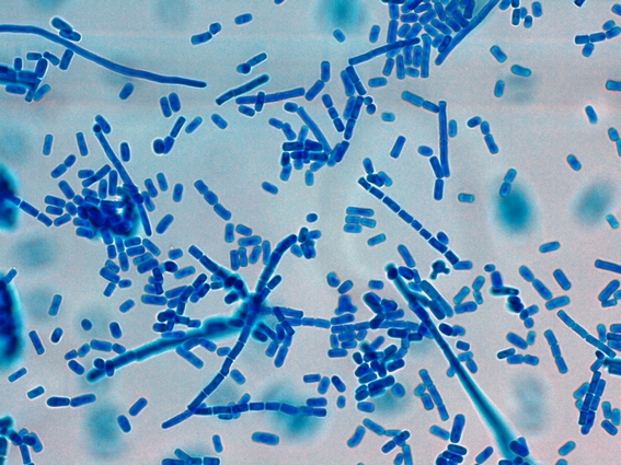
Hyphae are hyaline (clear/non-pigmented) and are septate (show divisions or walls within the hyphae).  Conidiophores are rather short (8 - 14 µm) and usually non-septate when compared to other Fusarium species. The conidiophores have a somewhat inflated appearance as their sides aren’t parallel but slightly bulge out slightly in the middle.  These conidiophores (phialides, or more accurately monophialides) are produced singly as they extend from the aerial mycelium.  Microconidia (5 - 12 X 2.3 – 3.5 µm) are usually non-septate, ellipsoidal and are straight or slightly curved in shape as they are abundantly produced from the tip of these phialides.  Microconidia are produced singly and never in chains.  These microconidia may accumulate around the tip of the phialide if not dispersed (false head).
Macroconidia (23-54 X 3.0 – 4.5 µm) are fusiform in shape (hence the genus name), and have a slightly pointed apical tip with a basal ‘foot’ cell (pedicellate) at the opposite end.  The fusiform macroconidia are also somewhat curved making it appear sickle-shaped or perhaps canoe-shaped in profile.  They usually contain 3-5 compartments or divisions within the macroconidium.
Smooth-walled terminal or intercalary chlamydospores (5 – 13 µm dia.) may be found.

 Fusarium oxysporum - First look at low power (X100 LPCB)

Fusarium oxysporum (X400 LPCB: DMD-108)

Fusarium oxysporum (X400 LPCB: Nikon)
Microconidia can be seen accumulating around the tips of the phialides (see below).

Fusarium oxysporum (same photo as above but cropped)
Arrows point to tips of phialides where microconidia are produced and accumulate

Fusarium oxysporum - Microconidia and a few Macroconidia (X100 LPCB: Nikon)

Fusarium oxysporum - again showing hyphae from which monophialides extend, producing microconidia at the tips which can accumulate there unless dispersed.
 (X1000 LPCB (DMD-108)

Fusarium oxysporum - another view (as above) - Note micro bar at top of photo.
 ( X1000 LPCB: DMD-108)

Fusarium oxysporum - yet another view of two parallel hyphae from which the phialides extend producing micro (&/or macro) conidia.  Conidia vary in shape from the rather straight fusiform (lens shaped) to the curved banana or canoe shape.
 (X100 LPCB: DMD-108)

Fusarium oxysporum - microconidia accumulating at the tips of phialdes
(X1000 LPCB: DMD-108)

Fusarium oxysporum - microconidia in various stages of development (arrows) at the tips of the monophialides from which they are produced.
 (X1000+10* LPCB: DMD-108)

Fusarium oxysporum - (yeah, I like photos) -again, microconidia accumulating around tips of phialides (X1000+10* LPCB: DMD-108)

Fusarium oxysporum - somewhat inflated (sides not parallel) phialides extending from hyphae where conida are produced (arrows).  The one on left has separated from the tip of the phialide whild the one on the right is young and still developing.
  (X1000+10* LPCB: DMD-108)

Fusarium oxysporum - micro & macro conidia accumulated along side of hyphae.
(X1000+10* LPCB: DMD-108)

Fusarium oxysporum - slightly curved micro & macro conidia pictured
(X1000+10* LPCB: DMD-108)

Fusarium oxysporum - chlamydospore present (arrow).  Difficult to see from the orientation but I believe this was an intercalary chlamydospore (growing between the hyphae and not a terminal chlamydospore at the end of a hyphae.
  (X1000+10* LPCB: DMD-108)

Fusarium species - Just for comparison of the shape.  The photo on the left is of micro (and a few macro) conidia produced by Fusarium oxysporum while the one on the right is of microconidia produced by Fusarium dimerum.  The F.dimerum is showing a greater curved "banana" or "canoe" shaped microconidia thatn the F.oxysporum. (Magnification not noted: Nikon for both)

Pathogenicity:
Infections with Fusarium species (fusariosis) may be localized or become disseminated.  Fusarium species are frequent agents of mycotic eye infections, particularly the cornea (keratomycosis, endopthalmitis).  They have also been implicated in onychomycosis (nail infections), catheter infections, peritonitis, sinusitis and septic arthritis.  As with many other fungi immunocompromised and neutropenic patients may be at greater risk.  Fusarium may contaminate stored grain where some species can produce potent mycotoxins.  Food prepared from these contaminated grains may cause illness on ingestion.  Fusarium species may also be found as laboratory contaminants but must not be dismissed outright without further investigation.

Differentiation:
Fusarium oxysporum can be differentiated from F.solani complex which produce thick, blunt macroconidia and long, narrow mono-phialides as well as numerous rough-walled chlamydospores.  The colonies, however, may appear brownish, particularly on Potato-Dextrose agar   F.oxysporum can be differentiated from F.verticillioides as this species has candle-shaped mono-phialides as well as baton-shaped microconidia in chains that are not easily disrupted.  Colonies however may appear purplish as those of F.oxysporumF.proliferatum also is similar in appearance however produces polyphialides (many phialides as opposed to the single phialide in monophialides) from which delicate chains of microconidia can extend.  Macroconidia are seldom seen in most F.verticilliodes or F.proliferatum species unless induced by special media or exposed to UV light.

*DMD-108 microscope/camera is capable of taking photos with 1000X optical magnification plus and additional 10% digital magnification.

Sunday, 10 June 2012

Paecilomyces lilacinus


Paecilomyces lilacinus (Fungus)

Ecology:  Wide-spread cosmopolitan saprobe of soil and decaying plant material.  Paecilomyces species have been implicated in the spoilage of food.  Has been isolated from cosmetics and some species of Paecilomyces parasitize insects.  Grows well at 30oC but growth is restricted at 37oC

Macroscopic Morphology:  As with many fungi, colour is influenced by the media it grows on.  Colonies often display a faint violet or mauve colouration (hence the name derived from lilac) which may develop to a reddish-grey tint.  The reverse is unremarkable.  Colonies are woolly to floccose and growth is moderately rapid, reaching 3 – 4 cm in about a week.


 Paecilomyces lilacinus on SAB ~2 weeks at 30oC - Note slight lilac (purplish) colour.

Microscopic Morphology:  P.lilacinus produces hyaline (clear), septate hyphae.  Conidiophores stipes (3 -4 µm wide) usually arise from submerged hyphae and can achieve lengths between 400 µm – 600 µm.  Branched conidiophores give rise to clusters of phialides.  The phialides have a broader base which tapers to form a long, narrow neck giving a delicate elegant appearance if compared to Penicillium.  The phialides tend to curve away from the central axis of the conidiophore.  Conidia (2.5 – 3.0 µm by 2.0 – 2.2 µm) form at the apical end of the phialides and are ellipsoidal (spindle) to fusiform in shape.  They arise in rather long divergent chains which are easily disrupted.

 P.lilacinus LPCB (X400 -Nikon)

Ditto


 More of the same in order to give a sense of how P.lilacinus appears.  Delicate brush-like appearance with phailides turning away (outward) from stipe). Kind of resembles Neptune's trident! Conidia in chains are copiously produced and easily disrupted.  I found no advantage with either adhesive tape preparations or slide cultures in preserving the appearance.  (LPCB X 400 Nikon)

 P.lilacinus - another view of conidiophores and chains of conidia (LPCB X1000 I believe, DMD-108)
P.lilacinus - I hate books that offer one B&W photo or a simple line drawing as the sole example of a particular fungus.  I find it difficult to get a real sense of the organism.  So I tend to overdo it in these posts by offering a number of views.  (LPCB X1000 Nikon)

P.lilacinus - showing a long chain of conidia which are delicate & easily disrupted
(X1000+10 DMD-108)

P.lilacinus - showd delicate, elongated, flask-shaped phailides bending away (splaying out) from stipe. (LPCB X100, DMD-108)

P.lilacinus - final view of phailides and chaining conidia.
 Size difference from previous photos is due to cropping.
(LPCB X1000 Nikon)

Pathology:  Often considered an environmental contaminant however should not be immediately dismissed as such.  P.lilicinus has been isolated from ocular infections such as keratitis, endopthalmitis and corneal ulcers.  Also has been implicated in bursitis, sinusitis, endocarditis, pulmonary infections, cutaneous infections, catheter related infections and even onychomycosis (nail infections).  Diabetic patients and immunocompromised patients may be at greater risk of infection.  P.lilacinus been successfully treated with ketoconazole.


*   *   *

Saturday, 2 June 2012

Diphyllobothrium latum (revisited)


Diphyllobothrium latum (Helmith/Cestode)

Note:  This post is entitled Diphyllobothrium latum "Revisited" as I posted a photo several years ago when a fixed film camera was the only tool in my arsenal.  I'm sure you'll find these new photos superior.  To visit the old post click below;

Known as ‘Fish Tapeworm’ or ‘Broad Tapeworm.  

Disease:  Causes’ Diphylobothriasis’.  In some instances the infection can be relatively harmless while in others it results in an illness resembling pernicious anemia.  The tapeworm may be depriving the infected host of vitamin B12 as the adult worm contains a high concentration of this vitamin.  Disease is acquired by eating raw or insufficiently cooked fish.  Campers who cook their catch of fish over an open fire in the wild and cultures who eat raw or pickled (incomplete) fish may be more likely to acquire the infection.  Lives in the proximal portion of the jejunum.

Geographic Distribution:  Found in temperate regions with cold clear lakes.  Prevalent in Japan, Russia, Scandinavia. Finland, the Baltics as well as Canada and Alaska.   Has been found in the Great Lake Regions of the United States however is not considered to be endemic.   In other regions of the world, different species of Diphyllobothrium may infect humans and susceptible mammals.

Life Cycle:  Diphyllobothrium latum required two intermediate hosts in order to complete its life cycle.  Briefly, the eggs are passed from feces which then hatch into small ciliated coracidium larvae.  These coracidia survive until they are ingested by copepods where the second larval stage (procercoid) develops with growth.  These fresh water crustaceans are themselves ingested by fish where they continue to grow where they develop into the plerocercoid stage.  Small infected fish may not be suitable for human consumption however they in turn may be ingested by larger fish thereby infecting them.  The larvae invades and resides in the flesh of the fish.  As the larger fish consumes smaller infected fish, the infectious potential increases when consumed by humans.  The plercoceroid larva (or sparganum) is not digested but remains in the small intestine where it develops to an adult tapeworm in about three to five weeks. The tapeworm, unless treated, may live for 25 years or longer.

Diagnosis: Infection with Diphyllobothrium latum is made by demonstrating the ova (eggs) in feces.  Occasionally segments of the proglottids (broken off segments of the worm) can also be found in the feces.

Ova (Eggs):  The Diphyllobothrium latum ova are ovoid in shape and have an operculum.  (a small cover or hatch through which the larvae can escape).  The yellowish-brown eggs are moderately thick-walled and are usually about 58 – 75 µm by 40 – 50 µm in size.  They may have a small know at the end opposite of the operculum however it may be indistinct.  The egg is unebryonated when passed in the feces.

Adult Worm:  The clinical laboratory usually encounters the egg rather than the adult worm as it is the stool (feces) which is usually submitted for diagnosis.   As I have no adult worm specimen, I’m unable to present any photos.  Consult other sources for photos of the worm which may grow to 4 – 10 meters in length.

 Two Diphyllobothrium latum eggs (arrows) in fecal concentrate (X100 Nikon)

 Two Diphyllobothrium latum eggs in fecal concentrate (X250 Nikon)

 Ditto

 Diphyllobothrium latum egg.  Arrows point to the the operculum or 'hatch' through which the larvae can escape. The protuberance, which can be somewhat inconspicuous, usually appears as a slight 'point' or 'bump' on the end opposite of the operculum. It is not evident on any of the photos in this post.  It does appear in my 2008 D.latum post. Fecal concentrate. Click on photo to enlarge for better viewing.
(X250 Nikon - size differs from previous photos due to cropping)

Broken Diphyllobothrium latum egg.  This is not an open operculum but rather a broken egg.
(X400 Nikon -Fecal Concentrate) Note micron bar in upper right.

Diphyllobothrium latum egg (X1000+10* -DMD-108: Fecal Concentrate)

Diphyllobothrium latum egg (X1000+10 DMD-108 Hematoxylin Stain)

Diphyllobothrium latum egg (X1000+10 DMD-108 Hematoxylin Stain)
(appearance differs due to perspective/orientation of egg)

Diphyllobothrium latum egg (X1000 DMD-108 Hematoxylin Stain)
(appearance differs due to perspective/orientation of egg)
Note micron bar in upper portion of photo

Diphyllobothrium latum Computer Screen Wallpaper (1024 X 768 when posted)
Ova in fecal concentrate X1000+10* DMD-108

*DMD-108 X1000 optical magnification (objective) + X10 digital magnification.


*   *   *

Saturday, 19 May 2012

Geotrichum candidum


Geotrichum candidum  (yeast/fungus)

Ecology: Ubiquitous worldwide distribution.  Has been isolated from soil, water, sewage, cereals, dairy products and various plants.   Geotrichum species have been considered a part of normal commensal flora when isolated from the sputa and/or feces of health humans.  The genus Geotrichum has several species with Geotrichum candidum being the most common.

Macroscopic Morphology:   On SAB, colonies exhibit moderately rapid growth, producing off-white to cream coloured colonies with a butyrous texture with a velvety, suede-like or ground glass/matt appearance.  Colonies grow best at around 25oC to 30oC however growth may be restricted at 37oC.
 Geotrichum candidum on SAB agar incubated at 30oC for 5 days

Microscopic Morphology:  Geotrichum species produce hyaline (clear), septate hyphae which show dichotomous branching (7µm – 11 µm wide).  Advancing undifferentiated aerial hyphae produce chains of arthroconida which fragment into individual cells of variable size (6 -12 µm to 3 – 6 µm).  Cells can be cylindrical in shape or may become barrel shaped.  Blastoconidia, conidiophores and pseudohyphae are not produced by Geotrichum species.  Disjunctor cells (empty cells in between arthroconida that fragment to release the arthroconidia) are absent which differentiates them from Coccidioides immitis and Malbranchea.  . Blastoconidia, conidiophores and pseudohyphae are also not produced.

Note: Photos taken with the Leica DMD-108 Microscope.  (a +10 after any magnification indicates another 10% digital magnification factor in addition to the optical magnification of the objective.

 Geotrichum candidum  at ~24 hrs (X250) LPCB

 Geotrichum candidum  at 48 hrs (X250) LPCB

 Geotrichum candidum   (X400) LPCB

Ditto

Geotrichum candidum showing hyphae, arthroconidia in chains and individual arthroconidia from the fragmentation of the chains  (X400) LPCB

Geotrichum candidum showing chains of arthroconida and separate arthroconidia
(X1000) LPCB

Geotrichum candidum showing chains of arthroconida and separate arthroconidia
(X1000+10) LPCB

 Geotrichum candidum showing chains of arthroconida, individual arthroconida from the fragmentation of the chains and septate hyaline branching hyphae in the lower right of photo.
(X1000+10) LPCB

Geotrichum candidum - yet another view to get a feel of the organism,
(X400) LPCB

Geotrichum candidum - showing hyaline hyphae with some rudimentary branching and evidence of the development of arthroconidia as chains towards the right.  Individual arthroconida appear throughout photo.  (X400) LPCB

Geotrichum candidum - individual arthroconidia.  Some of the arthroconidia appear as cylindrical with rounded ends.  Some arthroconidia look somewhat barrel-shaped with some variation in size.  Unsure of the round cell near the middle (X1000+10) LPCB

 Pathogenicity:  Geotrichum species is the causative agent of geotrichosis.  Broncheal and pulmonary infections are the most frequently reported form of the disease, particularly in the immunocompromised host.  Oral, vaginal, cutaneous and alimentary infections have also been reported.

Geotrichum species (candidum) showing dichotomous branching and chains of arthroconida which are fragmenting into individual arthroconidia.  Some cells appear with rather square ends and some rather round.  (X1000+10) LPCB
This photo intended as computer wallpaper (1024 X 768) when posted.


Sphaerita spp.


Sphaerita spp.

“Big bugs have little bugs, on their backs to bit them’
Little bugs have lesser bugs, and so on ad infinitum”
Anonymous

Some protozoan parasites can themselves be parasitized.  A hyper-parasite! The genus Sphaerita is considered to be a lower fungus and some species are capable of invading the cytoplasm of some amoeboid parasites.  Another parasite of parasites is Nucleophaga species which invades the nucleus.  Sphaerita, (sometimes called Polyphaga spp.) appear as tightly packed clusters within the cytoplasm and measure approximately 0.5 µm to 1.0 µm.

The parasite show below is possibly an Entamoeba coli, however the nucleus is not visible as it is out of the plane of focus.  Sphaerita appears as the small dots within the cytoplasm.

 An amoeboid parasite itself parasitized by the fungal parasite Sphaerita spp.  The Sphaerita appear as the neatly arranged circular dots withing the cytoplasm.
 Iron hematoxylin stain (X1000) Nikon


 An amoeboid parasite itself parasitized by the fungal parasite Sphaerita spp.  Doesn't appear to be digested material or inclusions. This parasite appears to have a rather dark blotchy karyosome reminiscent of Iodamoeba  bütchlii.  Unfortunately I failed to record measurements when I took these photos.  E.coli, Iodamoeba  bütchlii and E.nana were all present in this particular specimen.
Iron hematoxylin stain (X1000) Nikon

  Return Home (most recent posts)