(Note: For Candida dubliniensis, scroll to bottom of this post.)
I thought I’d start off my return from ‘sick leave’ by checking out the lab’s new digital camera. Just back, I have no “exotic” isolates to play with and explore so I’ve just retrieved a common, everyday Candida albicans. I’ve often, been disappointed in photographs found in most textbooks as they are small in size, black & white, poorly focused and often scrunched between a few lines of description. This yeast deserves a bit more respect as it is the most common cause of candidiasis, an infection which can range from acute, sub-acute or chronic. It may be present, yet unrecognized or have devastating, life threatening consequences. C.albicans may be present as commensal flora in the normal mouth, the skin and in the stool. Problems arise when, for what ever reason, the microbial balance is upset and Candida is allowed to overgrow other organisms which may assist in keep it in check. Antibiotic therapy may lessen normal bacterial flora allowing Candida to flourish. A change in the pH of the healthy vagina may result in a yeast infection. Immunosuppressive therapy may predispose a patient to infections by yeast.
Macroscopic Morphology:
Candida albicans grows rapidly in culture, reaching maturity in as little as three days. Colonies are cream coloured, raised, entire, smooth & butyrous. On enriched media such as Blood Agar, or Chocolate Agar, the colonies may develop small striations or outgrowths often referred to as “feet” which are indicative of the Candida albicans species.
 Candida albicans on Sabouraud-Dextrose Agar at 48 hours at 30C
(click on photo and illustration at right to enlarge for better viewing)
 Microscopic Morphology:
 A smear made from colonies taken from Sabouraud-Dextrose Agar, or blood agar will appear as round to oval cells about 4 to 8 µm. Though the cell wall structure differs from that of gram positive bacteria, yeast cells retain the crystal-violet stain of the routine gram stain and therefore appear purple. The yeast cell divides by budding. On primary media (reduced nutritionally) the budding can create elongated cells which when lined up along the dividing plane, mimic the appearance of a hyphae however these inline individual cells are referred to as a pseudohyphae (false hyphae). Some true hyphae may also be formed.
Along side of the pseudohyphae, Candida albicans develops blastoconidia around the area of the ‘septa’ (division). These appear as smaller round ‘grape-like’ clusters.
* * *
 Candida albicans retaining crystal violet stain from routine gram stain taken from SAB Agar
(click on photos to enlarge for better viewing) X1000
 Refractile Chlamydospore production on Corn Meal Agar at 2 hrs at 30C
(click on photo to enlarge for better viewing)
Technique: Chlamydospores production is best induced using the Corn Meal Agar (CMA) or Oxgall Agar. Inoculate the plate by picking up a sample of the Candida albicans colony with a straight wire and scratching the surface of the agar with it. With the surface inoculated, cover the scratched area with a microscope cover slip. This has a two-fold purpose. The glass cover slip reduces the atmospheric tension under the surface and protects the microscope objective from damage when viewing the plate after incubation. A filter paper moistened with sterile water and incubation at room temperature in the dark, may further encourage the production of chlamydospores After incubation (24-48 hours) remove the petrie dish plate cover (and microscope stage if you wish) and view the growth by carefully lowering the objective over the cover-slipped agar (X100-250). Viewing the growth around the edge of the glass cover slip should yield the best results.
 Refractile Chlamydospores and smaller 'grape-like' Blastoconidia (Blastospores) produced on Oxgal Agar and photographed through the microscope cover slip with the agar plate placed on the microscope stage.
(click on photo to enlarge for better viewing)
Yet another characteristic of Candida albicans is that it has the ability to produce 'Germ Tubes' when placed in a nutritionally rich horse serum. Inoculate about 1 to 2 ml of horse serum and incubate at 37C for about 2 to 3 hours (too long may result in the formation of pseudohyphae, mimicking germ tubes) . Examine under the light microscope for a protrusion growing out from the yeast cell. Germ tubes appear as outgrowths from the side of the yeast cell and although characteristic of Candida albicans, be aware that the closely related Candida dublinensis can also produce germ tubes. Other characteristics which won't be discussed here can easily be used to separate these two species. Germ tubes protrude from the originating cell with no "pinching" seen at the point where they extend. If there is evidence of pinching, this may not be a germ tube but rather the beginning pseudohyphal growth.
 Candida albicans germ tube production in Horse Serum at 37C after 3 hours incubation.
Note; there is no constriction/pinching where the germ tube leaves the originating cell indicating that this is indeed a germ tube and not the beginning of a pseudohyphae. (wet prep X400)
(click on photo to enlarge for better viewing)
Below are a few photos of Candida albicans as seen in actual specimens;
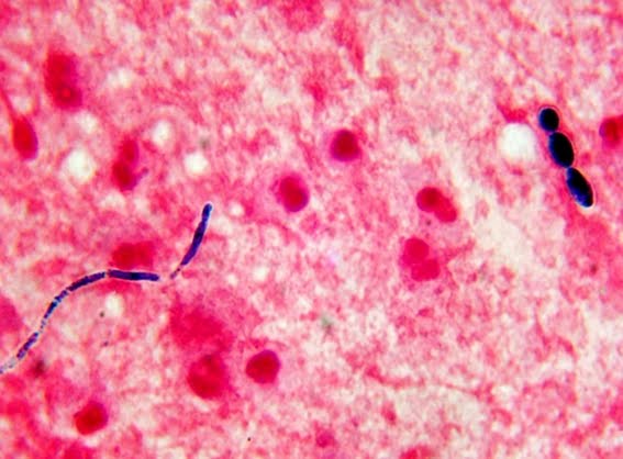
 Candida albicans in a vaginal swab seen in the form of a pseudohyphae and a few individual oval cells. (Gram Stain X1000)
(click on photo to enlarge for better viewing)
 Candida albicans seen as a pseudohyphae at lower left and individual yeast cells at upper right.
(Gram Stain of Sputum specimen X1000)
(click on photo to enlarge for better viewing)
 Candida albicans pseudohyphae and individual cells seen in an aspirate from a kidney.
Depending of various factors, individual yeast cells, pseudohyphae, or both may be present in any given specimen.
(click on photo to enlarge for better viewing)
New: April 01, 2012
An interesting photo of a fungus as it appeared in a direct blood culture gram stain. Identification proved it to be Candida albicans. Just had to share my beautifully bizarre photo - hard to imagine this spiny ball of spikes bouncing around inside someones blood vessels!
 Candida albicans in direct smear of Positive Blood Culture.
(Usually seen as the round to oval cellular form and not as these clumps of pseudohyphae)
* * *
New: April 18th, 2015
Candida albicans vs Candida dubliniensis
Okay, you think you have isolated Candida albicans. But is it
really?....and why should you care?
Well, Candida albicans & Candida dubliniensis are virtually indistinguishable
using common physiological tests (see list which follows). However, there may be one significant
difference which should be considered. Candida dubliniensis may exhibit increased
resistance to the anti-fungal agent Fluconazole.
This is of particular concern when this species is isolated from sterile
sites such as blood cultures where delayed or failure of treatment may be
fatal.
• Both yeasts are virtually identical in their physiological characteristics
• Carbohydrate assimilation is virtually identical ( some variation +/- vs -/+ of little use in identification or differentiation)
• Glucose fermentation – both positive
• Chlamydospore production – both positive
• Germ tube production – both positive
• Urease – both negative
• Nitrate – both negative
• Cyclohexamide (0.04%) – both positive for growth.
• Growth at 30oC & 37oC -both positive
While some commercial identification systems claim they can
distinguish between the two species, this is a costlier solution to a problem
that can be solved using incubation temperature alone.


C.dubliniensis
• Growth at 42oC to 45oC = -/w
• Fluconazole –may exhibit increased resistance.
C.albicans
• Growth at 42oC to 45oC = +/-
• Fluconazole – generally considered susceptible.
• Growth at 42oC to 45oC = -/w
• Fluconazole –may exhibit increased resistance.
C.albicans
• Growth at 42oC to 45oC = +/-
• Fluconazole – generally considered susceptible.

.jpg)


