Pithomyces species -Hyphomycete
Ecology:
The
genus Pithomyces has approximately 50
recognized species to date. Speciation
is most accurately achieved by molecular means; however, careful observation of
morphological features can identify this mould to the genus level.
Pithomyces species are dematiaceous
saprobes (darkly pigmented moulds which commonly grow on dead organic matter)
and may be found on the leaves and stems of a variety of plants. They have also
been isolated from decaying wood, tree bark (Acacia) and from soil.
Pathogenicity:
Pithomyces species have been
implicated in human disease however their role has not been sufficiently
substantiated. Pithomyces has reportedly been isolated
from finger and toe nails, a hand lesion (skin scrapings), peritoneal fluid, bronchial
washings, and from a chronic nasal polyposis. The mould may also contribute to
general allergic reactions. In the
United States, the most commonly isolated Pithomyces
species appear to be P. chartarum, P. sacchari, and P. maydicus.
Pithomyces species have been
implicated in pithomycotoxicosis (facial eczema) of ruminants such as sheep,
cattle and goats. P.chartarum, in particular is considered the cause of facial eczema
in sheep.
Pithomyces species are commonly
considered to be laboratory contaminants, however, they should not be ruled out
without careful consideration, particularly in immunocompromised patients.
Macroscopic Morphology:
Pithomyces exhibits fairly rapid
growth, maturing in about five days to a week.
Colonies on SAB at 30ᵒC are olivaceous, light to dark brown to
brownish-black. Colour is species and
media dependant. Dark brown to black
areas, may be seen macroscopically on some species (P. atro-olivaceus), revealing sporodochia (pleural of
sporodochium), which are areas of greater conidial production. The overall colonial texture is downy to
cottony, with a short feathery nap (effuse).
The reverse is brown to brownish-black in colour.
The
isolate presented in this blog (SAB 30ᵒC) post had a lighter cream coloured
fringe or edge to the colony.

Pithomyces species - Sabouraud-Dextrose Agar (SAB), 1 Week, 30ᵒC (Nikon)
Microscopic Morphology:

Pithomyces species produce septate,
sub-hyaline (pale to light brown) hyphae.
Conidiophores are generally short (peg-like, ~10 µm length), and rather
poorly differentiated from the vegetative hyphae from which they extend. Conidia (10 – 17 µm X 18 – 30 µm) are
produced singly at the apex of the conidiophore where they are attached by a
short denticle. After conidial
dehiscence (release of conidia), a visible annular frill may remain at the
conidial base where once attached to the conidiophore. Conidia are muriform (have both longitudinal
and transverse septations) and are broadly ellipsoidal to ovate (egg shaped) or
pyriform (pear shaped) in shape. P.chartarum usually exhibits 2 – 5
transverse septa with 0 – 3 longitudinal septa. The muriform or septation
pattern may be species dependant; P.
atro-olivaceus may only produce horizontal septa. Conidia are dark brown in colour when mature
and usually have an echinulate (spiny or prickly) or verruculose/verrucose (warty)
texture.
Caution: Micron scale (µm) may change between 50 or 100 µm at higher magnifications.
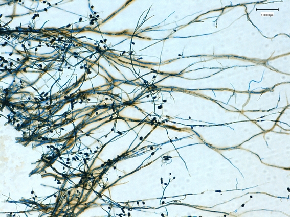
Pithomyces species - Initial view -growth from the edge of a slide culture.
(250X, LPCB, DMD-108)

Pithomyces species - darkly pigmented conidia with internal septations are seen.
(400X, LPCB, DMD-108)

Pithomyces species - Numerous, pigmented conidia seen. Insert shows the muriform (both longitudinal and transverse septa) septations.
(400X, LPCB, DMD-108)

Pithomyces species - after conidial dehiscence (release of conidia), a visible annular frill may remain at the conidial base where once attached to the conidiophore. (400X, LPCB, DMD-108)

Pithomyces species - conidia are broadly ellipsoidal to ovate (egg shaped) or pyriform (pear shaped) in shape. (400X, LPCB, 400X)

Pithomyces species - Conidia are borne on short stalks. Brownish pigment has exuded from the hyphae and can be seen as the brown haze alongside the hypha. (400X, LPCB, DMD-108)

Pithomyces species - conidiophores are generally short (peg-like, ~10 µm length), and rather poorly differentiated from the vegetative hyphae from which they extend. (400X, LPCB, DMD-108)

Pithomyces species - as above. (400+10X, LPCB, DMD-108)

Pithomyces species - conidia (10 – 17 µm X 18 – 30 µm) are produced singly at the apex of the conidiophore where they are attached by a short denticle. (400+10X, LPCB, DMD-108)

Pithomyces species - septate hypha with pigment seen along the outer walls of several. Conidium on short stalk is seen at center right. (400+10X, LPCB, DMD-108)

Pithomyces species - single, elongated conidium seen at the apex of a withering hyphal element or conidiophore. (400+10X, LPCB, DMD-108)

Pithomyces species - some chains appeared to be formed by this particulate isolate. Only one source I consulted (Davone -see sidebar) stated that Pithomyces species do not chain. I isolate presented here conforms to the characteristics described for Pithomyces with the exception of chain formation by the conidia. This should not be confused with the chain-like formation of conidia as seen in Alternaria species. (400+10X, LPCB, DMD-108)

Pithomyces species - ditto.
(400+10X, LPCB, DMD-108)

Pithomyces species - yet another photo at higher magnification...
(1000X, LPCB, DMD-108)
(1000X, LPCB, DMD-108)

Pithomyces species - oval conidium at apex of a short stock which shows little differentiation from the vegetative hyphae. (1000X, LPCB, DMD-108)

Pithomyces species - pigment escaping from the hyphae into the surrounding medium.
Vegetative mycelium composed of thin-walled hyaline, septate, smooth or verrucose, septate hyphae, 4–7 µm diameter, which may give rise to chains of verrucose, one-celled, dark brown, intercalary chlamydospores 10 -20 µm X 8 - 18 µm[i].
Vegetative mycelium composed of thin-walled hyaline, septate, smooth or verrucose, septate hyphae, 4–7 µm diameter, which may give rise to chains of verrucose, one-celled, dark brown, intercalary chlamydospores 10 -20 µm X 8 - 18 µm[i].

Pithomyces species - pigment escaping from the septate hyphae into the surrounding medium. Annular frill can be seen attached to the anterior end of the conidium.
(1000X, LPCB, DMD-108)
(1000X, LPCB, DMD-108)

Pithomyces species - more intercalary chlamydospores seen as described two photos above.
(1000X, LPCB, DMD-108)
(1000X, LPCB, DMD-108)

Pithomyces species - Conidia are dark brown in colour when mature
and usually have an echinulate (spiny or prickly) or verruculose/verrucose (warty)
texture. The conidium at center-right clearly shows a spiny or prickly surface. Intense uptake of the Lactophenol Cotton Blue (LPCB) dye somewhat obscures the muriform septations within the conidium.

Pithomyces species - appears to be attached at both ends which would make it an intercalary chlamydospore (?)
(1000+10X, LPCB, DMD-108)
(1000+10X, LPCB, DMD-108)

Pithomyces species - short, peg-like, conidiophores arising from the vegetative hyphae at right-angles with single pigmented, muriform conidium at each apex.
(1000X, LPCB, DMD-108)
(1000X, LPCB, DMD-108)

Pithomyces species -here you get it all as described in previous photos. Pigmented septate hyphae with pigment escaping into the surrounding medium. Prickly surfaced muriform conidia borne singly on short peg-like conidiophores. The free conidium closest to the top shows the annular frill which remains from where it was attached to the conidiophore.
(1000X, LPCB, DMD-108)
(1000X, LPCB, DMD-108)

Pithomyces species -two, rather smooth walled, conidia attached to the hypha by short conidiophores.
(1000+10X, LPCB, DMD-108)
(1000+10X, LPCB, DMD-108)

Pithomyces species - some chaining (?) evident.
(1000X, LPCB, DMD-108)
(1000X, LPCB, DMD-108)

Pithomyces species - single muriform conidium at the end of a short conidiophore.
(1000+10X, LPCB, DMD-108)
(1000+10X, LPCB, DMD-108)

Pithomyces species - single muriform conidium at the end of a short, rather twisted, conidiophore.(1000+10X, LPCB, DMD-108)

Pithomyces species - conidia are borne singly at the ends of the conidiophores.
(1000+10X, LPCB, DMD-108)
(1000+10X, LPCB, DMD-108)

Pithomyces species - I'm trying to figure this one out. Are those two conidia arising from two, obscured, conidiophores, or is one an intercallary chlamydospore with a conidiophore & conidium arising from the same area of the hypha?.

Pithomyces species - Sources state that Pithomyces conidiophores produce single conidia. Does this photo show a single conidium at the end of a short, pale pigmented conidiophore or is conidiphore the LPCB stained structure arising from the hyphae below with the apex of the conidiophore branched, and one conidium missing? You decide...

Pithomyces species -muriform conidia at the end of short peg-like conidiophores.
(1000+10X, LPCB, DMD-108)
(1000+10X, LPCB, DMD-108)

Pithomyces species -conidia texture described as echinulate (spiny or prickly) or verruculose/verrucose (warty)
texture.
(1000+10X, LPCB, DMD-108)
(1000+10X, LPCB, DMD-108)

Pithomyces species - conidia which aren't over-saturated with the LPCB stain and more clearly show the muriform septation within.

Pithomyces species - thickened and roughened wall of the intercalary chlamydospores.
(1000+10X, LPCB, DMD-108)
(1000+10X, LPCB, DMD-108)

Pithomyces species - chaining of conidia (?) at left. Two, rather young conidia on short peg-like conidiophores at lower center of photo.
(1000+10X, LPCB, DMD-108)
(1000+10X, LPCB, DMD-108)

Pithomyces species
(1000+10X, LPCB, DMD-108)
(1000+10X, LPCB, DMD-108)
Pithomyces species have to be differentiated from closely
related dematiaceous hyphomycetes such as Ulocladium
species, Stemphylium species, Alternaria species and Epicoccum species. (See Table Below)

Too small to read? Click on table to get image. Now right click on image and select 'view image'. In Windows, cursor now has a + sign within it. Click on image of table to now magnify the table.
Alternatively, just click and download the damn thing...
[i] The
Genus Pithomyces in South Africa
W.F.O. Marasas and Ingrid H. Schumann,
Bothalia 10, 4:
509 – 516, 1972
* * *


.jpg)



















