Ascaris lumbricoides (Intestinal
Nematode) “Roundworm”
Geographic
Distribution:
Ascaris
lumbricoides can be found throughout the world but is more commonly found
in moist temperate and tropical regions.
Ascaris is increasingly
prevalent where poor sanitation exists.
In cultures where human excrement (night soil) is used as fertilizer,
infection may again be greater.
Pathogenicity:
Infection occurs with the ingestion of fully embryonated
eggs (ova). Infection with Ascaris lumbricoides is termed
Ascariasis and it affects more of the world’s population than any other
parasitic disease. Ingestion of small numbers of eggs may be
asymptomatic for the host however larger numbers may cause Ascaris pneumonitis[i]
or Loeffler’s[ii]
syndrome. Those infected with Ascaris lumbricoides may develop
asthmatic attacks, shortness of breath, wheezing or a persistent cough. A heavy infection in the small intestine may
result in a bowel obstruction, especially in children. Symptoms may include fever and generalized
malaise possibly with abdominal distension, associated tenderness and possible
vomiting.
Life Cycle:
 Ingested embryonated eggs travel to the duodenum (small
intestine) where they hatch and begin an obligatory migration throughout the
body before they return to the duodenum to mature into adulthood. Hatched larvae start the migration by
penetrating the duodenum wall to enter the blood or lymphatic system which
eventually carries the larvae to the liver and the heart to eventually enter
pulmonary circulation. In the lungs, the
larvae break free of the capillaries to enter the aveoli where they continue to
grow. After about 3 weeks, they begin to
migrate through respiratory passages to reach the esophagus where they are
swallowed and once again reach the small intestine.
Ingested embryonated eggs travel to the duodenum (small
intestine) where they hatch and begin an obligatory migration throughout the
body before they return to the duodenum to mature into adulthood. Hatched larvae start the migration by
penetrating the duodenum wall to enter the blood or lymphatic system which
eventually carries the larvae to the liver and the heart to eventually enter
pulmonary circulation. In the lungs, the
larvae break free of the capillaries to enter the aveoli where they continue to
grow. After about 3 weeks, they begin to
migrate through respiratory passages to reach the esophagus where they are
swallowed and once again reach the small intestine.
At about two to three months post ingestion of the eggs,
the now mature worms begin to lay their own eggs. It has been estimated that mature female
worms may produce an average of 200,000 eggs daily. Ascaris lumbricoides females are oviparous[iii]. When passed, the eggs require about 2 to 3
weeks outside of the host to develop into the infective embryonated stage. While the eggs are susceptible to excessive
heat and drying, they can remain viable in moist soils for long periods of
time.
Ascaris Worm:
The adult Ascaris lumbricoides worms area creamy-white in
colour, occasionally with a pinkish cast.
Female worms range from 20 to 35 cm in length. Males are generally shorter and usually do
not exceed 30 cm. Female worms are also
generally thicker (3-6 mm) than the more slender males (2-4 mm). Males can be distinguished from females by
their incurved tail. Females have a
straight tail. Adult worms are believed
to live up to a year. Ascaris worms do
not attach themselves to the intestinal wall but rather maintain their position
through constant movement.
.jpg)
Ascaris lumbricoides: Adult male worm show with its distinctive curved tail. This particular worm was about 24 cm or 10 in in length, (Nikon-Macroscopic)
Ascaris Eggs (Ova):
Diagnosis of ascariasis is by the demonstration of the
characteristic eggs in the feces.
Fertile eggs
are broadly oval in shape and typically yellow-brown in colour, stained by the
bile in freshly passed stools. They
measure 45 to 75 µm in length by 35 to 50 µm in breadth. The outer albuminoid coat is coarsely
mammillated covers a smooth shell which is difficult to distinguish in
laboratory preparations. Some confusion
in diagnosis may occur if the Ascaris
egg has lost its mammillated coat (decorticated) as it may somewhat resemble Hookworm or Trichostrongylus species
ova. Ascaris eggs contain only one cell
when passed in the feces.
Infertile eggs are
elongate, about 85 – 95 µm by 43 – 47 µm in size. Their mammillated layer may vary from being
coarsely irregular to a relatively smooth layer, almost devoid of
mammillations. The internal contents of infertile eggs appear as a mass of
disorganized, refractile granules.
Note: All the following photos are take from the re-suspended sediment after fecal concentration.

Ascaris lumbricoides egg seen in the center of the photograph.

Ascaris lumbricoides egg has a distinctive appearance. This egg shows the rough looking surface due to the mammillated albuminoid coat.
(DMD-108, 400X)

Ascaris lumbricoides: another photo of the egg with its rough mammillated coat.
(Nikon, 400X)

Ascaris lumbricoides: a photo of the egg with an alternate focus inserted for comparison. As the egg is three-dimensional, adjusting the focus can aid in visualizing the egg's structure.
(Nikon, 400X)

Ascaris lumbricoides: as above.
(DMD-108, 400X)

Ascaris lumbricoides: Two A.lumbricoides eggs seen in the same field.
(Nikon, 400X)

Ascaris lumbricoides: Typical appearance of the Ascaris egg. The presence of these eggs in a fecal specimen is diagnostic for an Ascaris infection.
(DMD-108, 400+10X)

Ascaris lumbricoides: The thick albuminoid mammillated layer is quite evident on this fertile Ascaris egg. (DMD-108, 400+10X)

Ascaris lumbricoides: As above with a slightly altered focus.
(DMD-108, 400+10X)

Ascaris lumbricoides: Another photo of the Ascaris egg in a fecal concentrate.
(DMD-108, 400+10X)

Ascaris lumbricoides: As above but with an alternate focus of this fertile Ascaris egg. I post these alternate focus photos in an attempt to demonstrate the three-dimensional nature of the egg and their complexity. (DMD-108, 400+10X)

Ascaris lumbricoides: Yet another example, 'cause more is better!.
(DMD-108, 400+10X)
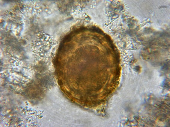
Ascaris lumbricoides: At a higher magnification. The large single cell interior is clearly visible.
(Nikon, 1000X)

Ascaris lumbricoides ova: Okay, more is better, right? Another photo showing the thick outer albuminoid mammillated layer. The inner wall often cannot be visualized unless the egg loses this outer layer (decorticated). The single celled contents are clearly visible.
(Nikon, 1000X)

Ascaris lumbricoides: Too many photos? I have to do something with them! Another photo of the Ascaris egg however it appears as if the interior cell has divided into four or more individual cells.
(Nikon, 1000X)

Ascaris lumbricoides: An Ascaris egg with the inset enlargement of the Ascaris egg. There appears to have been a number of divisions of the original cell contained within the egg.
(Nikon, 250X)
Too many photos? Just a few more to go..
While the Ascaris lumbricoides ova can be found in iron-hematoxylin stained preparations, the eggs usually stain so intensely that they appear as silhouettes and very little structural information can be discerned. They may be missed by an inexperienced eye. A thinner part of the stained slide may offer the best chance of recognizing and finding these eggs. The sedimentation concentration wet preparation technique is preferable in visualizing these eggs when present.
Note: The following photos are taken from Iron-Hematoxylin stained fecal preparations.

Ascaris lumbricoides:Two Ascaris eggs are seen in this photo which appear as two large dark masses on either side of the picture. The dark blob in the lower center is just an artifact present in the stool and clearly does not have the outline of the Ascaris egg.
(Nikon, 250X)

Ascaris lumbricoides: As with the concentrates, if a good microscopic field is found in the stained preparation, the Ascaris egg exhibits different textures as one focuses through it.
(Nikon, 1000X)

Ascaris lumbricoides: Same photo as above but with altered focusing. The mammillated layer appears as a scalloped edge surrounding the single celled contents of the egg.
(Nikon, 1000X)

Ascaris lumbricoides: In most iron-hematoxylin stained preparations the Ascaris ova will be heavily stained and often appear in silhouette with very little structure revealed.
(Nikon, 1000X)

Ascaris lumbricoides: As above. If a thin field can be found, the stained ova may show some detail.
(Nikon, 1000X)

Ascaris lumbricoides: Okay, last one. This egg doesn`t look particularly healthy. The `scalloped`edge of the mammillated coat is somewhat visible along the upper edge.
(Nikon, 1000X)
Diagnostic
Problems:
Both fertile and infertile Ascaris eggs concentrate well
however well using sedimentation techniques, however, if the laboratory only
uses a flotation method for concentrating, infertile eggs may be missed by this technique.
[i]
Pneumonitis refers to an inflammation of the lungs which can be cause by a
variety of agents, from allergic, chemical or an infection. Ascaris
pneumonitis is an inflammation caused by the presence of Ascaris
lumbricoides migration through the lungs.
[ii]
Löffler's syndrome or Loeffler's syndrome is a disease in which eosinophils
accumulate in the lung in response to a parasitic infection.


.jpg)


.jpg)



















