Eurotium herbariorum (Aspergillus glaucus)
Mould
Note: This fungus has both a sexual and asexual
means of reproduction. When both are
present, the sexual stage (teleomorph) name takes precedence over the asexual
(allomorph) name. Applied here, Eurotium herbariorum take precedence
over Aspergillus glaucus. However, I will generally refer to the fungus here as Aspergillus glaucus as most references
still continue to do so.
Ecology:
Aspergillus glaucus is a cosmopolitan fungus (worldwide distribution) and while
it prefers drier environments, it can be isolated from soils, house dust, plants
& dried foods. A.glaucus is also osmophilic, meaning it can grow, and perhaps
prefers to grow in environments containing a higher sugar concentration. Growth is restricted or limited at 35˚C which
may account for its limited pathogenicity.
Pathology:
A.glaucus is not very invasive and is rarely
encountered in the clinical laboratory.
It has been implicated as a cause of ocular (eye) infections,
particularly after some traumatic injury.
Cerebral, orofacial, cardiovascular and pulmonary infections are rare
but have been reported. May also cause
sinusitis (nasal) and otitis (ear) infections.
May be considered an opportunistic fungus particularly with
immunocompromised patients.
Macroscopic
Morphology:
Growth is slow to moderate, maturing in about 7 to 21
days. Colony size expands rather slowly.
Colony colouration is media dependent but is described as
a dull to deep green to a greyish turquoise, with yellow to orange areas where
cleistothecia are being produced. The
reverse is pale yellow to yellow. The isolate presented below failed to grow at 30˚C but grew well at ambient room temperature (~20˚C).

Eurotium herbariorum (Aspergillus glaucus) -SAB + ~20% Sucrose, Room Temperature, 14 Days
(this particular isolate failed to grow at 30˚C)
 Note on Culture: Our medical laboratory purchases all media from a commercial supplier and in these financially challenging times we stock only what can be clinically justified. We have no media room where we can concoct media from basic ingredients. Sabouraud Dextrose media has a sugar content (dextrose) of 4% which would favour the production of asexual conidia while a high osmotic concentration (~20%) would enhance cleistothecia production. In order to enhance growth and increase the production of cleistothecia, I obtained a couple of tubes of Sabouraud Dextrose (slants) and melted them down in a boiling waterbath. I did a rough 'back of an envelope' calculation of how much sugar I would have to add to bring the concentration up to about 20%. With no lab supply of dextrose to add, I went down to our hospital 'Tim Horton'™ coffee shop (a Canadian franchise) and obtained a packet of sucrose. Adding this to the melted SAB agar, I brought the total sugar concentration up to about 20% (glucose + sucrose) to raise the osmotic concentration. I sacrificed a sterile media petrie dish contents (as we also do not stock sterile empty petrie dishes) and poured in my own concoction of "Tim Horton's Cleistothecia Enhancing Media", or THCEM for short. For this reason you will see the media identification on the reverse plate blurred out. With no autoclave on site, I hoped that the boiled contents remained sterile and the high osmotic concentration would discourage other organisms from growing. The THCEM media worked extremely well! The photographs which follow are from this agar media.
Microscopic
Morphology:
Hyphae are septate and hyaline.
Teleomorph –
Sexual state is seen with the production of cleistothecia (ascomata). These structures are globose to subglobose,
about 60 µm to 150 µm in diameter. In
their natural state they appear yellow to golden in colour and their presence
may be seen macroscopically as distinctly yellowish areas within the maturing
colony. Within the
cleistothecia/ascomata, 8-celled asci are produced which are released at
maturity or when ruptured. The 8-celled
asci (10 µm - 12µm diameter) are dehiscent (dissolve) and release individual
ascospores on maturity). The Ascospores themselves
mature in about two weeks’ time and are lenticular (lens shaped) with a
noticeable longitudinal furrow. (On the
side, they resemble a hamburger, with the patty being the furrow.) They range between 5 µm to 7 µm by 3 µm to 5
µm in size). The ascospores have a
rather smooth surface texture which may help differentiate them from the
ascospores produced by the Neosartorya
species.


Allomorph – smooth walled conidiophores extend between 300 µm – 700 µm in length and are between 7 µm – 12 µm in width. Vesicles are globose (spherical) to subglobose (subspherical) to pyriform (tear-drop) in shape and roughly 18-30 µm in diameter. A.glaucus is uniseriate with phialides 7 – 11 µm to 3 – 7 µm in size and generally covers most of the vesicle. The conidia (4 µm to 8 µm diameter) are spherical to ellipsoidal in shape and are echinulate to spinose (finely roughened/fine spines).
Teleomorph (Eurotium herbariorum) - Sexual State

Eurotium herbariorum (A.glaucus) - a first look with an adhesive tape preparation from the colony. E.herbariorum produces numerous cleistothecia and production can be enhanced on media with a higher sugar content. Cleistothecia production on the plate can be seen macroscopically as an enhanced yellowish band within the colony.
(250X, KOH, DMD-108)
KOH= Potassium hydroxide

Eurotium herbariorum (A.glaucus) -cleistothecia at a higher magnification. The KOH kills the fungus so that the slide can be safely removed from the biological safety cabinet without fear of contamination. It also clarifies the preparation to some degree and dose not alter the natural colour of the mould.
(400X, KOH, DMD-108)

Eurotium herbariorum (A.glaucus) -as above.
(400X, KOH, DMD-108)

Eurotium herbariorum (A.glaucus) -The cleistothecia vary in size as they mature but are generally in the range of 60 µm to 150 µm in diameter. This size also aids in distinguishing Eurotium from other cleistotheia producing moulds.
(400+10X, KOH, DMD-108)

Eurotium herbariorum (A.glaucus) -the Aspergillus conidiophore (in rather poor shape) is seen on the left while a cleistothecium (also breaking up) is seen on the right.
(400X, KOH, DMD-108)

Eurotium herbariorum (A.glaucus) - A cleistothecium breaking apart and releasing ascospores. The KOH does not react with the contents of the cleistothecium as it did with Emericella nidulans (Aspergillus nidulans) where the contents produced a purple colour.
(400X, KOH, DMD-108)

Eurotium herbariorum (A.glaucus) -Several cleistothecia seen with the released asci staining blue with the LPCB.
(400X, LPCB, Nikon)

Eurotium herbariorum (A.glaucus) -a cleistothecium showing a break at the bottom and the asci within staining blue.
(400+10X, LPCB, DMD-108)

Eurotium herbariorum (A.glaucus) -another view as above. A released, intact 8-celled ascus can be seen on the bottom edge of the cleistothecium (or ascomata)
(1000X, LPCB, DMD-108)

Eurotium herbariorum (A.glaucus) -another view of a cleistothecium (Ascomata) with an 8-celled ascus inside. (1000X, LPCB, Nikon)

Eurotium herbariorum (A.glaucus) -two cleistothecia with the large one filling most of the upper right of the photo, filled with asci & apparently free ascospores.
(1000X, LPCB, Nikon)

Eurotium herbariorum (A.glaucus) -yet another view showing several 8-celled asci packets that have been released from the cleistothecia (Ascomata).
(1000X, LPCB, Nikon)

Eurotium herbariorum (A.glaucus) - a cleistothecium filled with asci.
(1000X, LPCB, Nikon)

Eurotium herbariorum (A.glaucus) -a free 8-celled ascus seen in the center of the photo with a cleistothecium on the right.
(1000X, LPCB, Nikon)

Eurotium herbariorum (A.glaucus) -two intact cleistothecia (ascomata) seen with a typical `cracked mud` appearance of the surface.
(1000X, LPCB, Nikon)

Eurotium herbariorum (A.glaucus) -A breach in the wall of a cleistothecium from where the asci contained within will escape.
(1000X, LPCB, DMD-108)

Eurotium herbariorum (A.glaucus) -a single, free 8-celled ascus seen in the center of the photo. This was released, with many others, when the cleistothecium matured and dissolved or broke apart. (1000X, LPCB, DMD-108)

Eurotium herbariorum (A.glaucus) -a slightly different appearance of a cleistothecium, with the point of attachement to the hyphae seen below it.
(400+10X, LPCB, DMD-108)

Eurotium herbariorum (A.glaucus) -two cleistothecia amongst the hyphae. The photo appears oversaturated with the blue of the LPCB masking some of the detail. I was unable to correct for this with using a photo editing program.
(1000X, LPCB, DMD-108)

Eurotium herbariorum (A.glaucus) -as above but here you now can see the rough-walled conidia which were produced by the Aspergillus glaucus allomorph.
(1000X, LPCB, DMD-108)
Allomorph – smooth walled conidiophores extend between 300 µm – 700 µm in length and are between 7 µm – 12 µm in width. Vesicles are globose (spherical) to subglobose (subspherical) to pyriform (tear-drop) in shape and roughly 18-30 µm in diameter. A.glaucus is uniseriate with phialides 7 – 11 µm to 3 – 7 µm in size and generally covers most of the vesicle. The conidia (4 µm to 8 µm diameter) are spherical to ellipsoidal in shape and are echinulate to spinose (finely roughened/fine spines).
Allomorph (Aspergillus glaucus) - Asexual State

Aspergillus glaucus (Eurotium herbariorum) -so here is the allomorph which is the asexual Aspergillus glaucus component. Both were present on this isolate.
(1000X, LPCB, DMD-108)

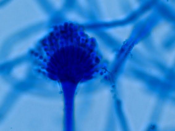
Aspergillus glaucus -a conidiophore and vesicle with the phialides bearing spiny, spiked or rough walled conidia. The DMD microscope somehow tends to over saturate in the photograph while the image on the high definition LED screen looks pristine. Again, I find that my attempts at correcting for this with photo editing programs falls short.
(1000X, LPCB, DMD-108)

Aspergillus glaucus -Here you can easily see that Aspergillus glaucus is uniseriate. If it were biseriate there would be an additional structure called a metulae between the phialide and the vesicle supporting it. (Kind of a double-decker structure). Again the rough, spiny conidia can be seen at the ends of of the phialides.
(1000X, LPCB, DMD-108)

Aspergillus glaucus -just `cause I like the photo. Vesicles
are globose (spherical) to subglobose (subspherical) to pyriform (tear-drop) in
shape and roughly 18-30 µm in diameter. Chains of conidia can be seen trailing from the apex of the phialides where they were produced.
(1000+10X, LPCB, DMD-108)

Aspergillus glaucus -a rather poor looking conidiophore and vesicle in the upper left however there is a very good example of the shape of the ascospores. Here you see the original 8-celled packet (ascus) somewhat broken apart but the equitorial trough (furrow) and two lateral crests are clearly visible (inset). The smooth texture helps to distinguish these ascospores from those produced by Neosartorya species. The kind of look like a hamburger patty within a bun as viewed from the side!
(1000X, LPCB, Nikon)

Aspergillus glaucus -here is a whole mess of A.glaucus conidia showing the spiny texture of the surface.
(1000+10X, LPCB, DMD-108)

Aspergillus glaucus -just to show the difference in size between the individual ascospores and the conidia produced by this species. Depending on the orientation in the field, the ascospores may not show the equitorial trough or crests. (ie viewed from the top and not the side)
(1000+10X, LPCB, DMD-108)

Aspergillus glaucus -one last photo of an Aspergillus glaucus conidium with its rough wall.
(1000X, LPCB, DMD-108)
Notes:
With the petrie dish under a stereoscopic dissecting microscope, the fruiting structures were both visible and distinguishable. The Aspergillus conidiophores stood erect like trees in a forest, with the conidia appearing a pale sage green in colour. The cleistothecia were interspersed and about the same size in total diameter but they appeared yellowish-brown in colour. A really beautiful sight viewed between 16X to 40X magnification!
With the petrie dish under a stereoscopic dissecting microscope, the fruiting structures were both visible and distinguishable. The Aspergillus conidiophores stood erect like trees in a forest, with the conidia appearing a pale sage green in colour. The cleistothecia were interspersed and about the same size in total diameter but they appeared yellowish-brown in colour. A really beautiful sight viewed between 16X to 40X magnification!
The Aspergillus
glaucus group contains approximately 17 species (subject to continued molecular study and reassignment, I’m sure). Of these, some species are extremely
xerophilic (need little or no water to survive) and as such, may be found in
greater numbers in tropical to subtropical regions as opposed to temperate
zones. This property also favours their
growth on dry or concentrated substances.
As mentioned earlier, they are osmophilic and can grow in the presence
of high sugar concentrations where the carbohydrate draws away the water. That fungus growing on top of the jam jar may
be Aspergillus glaucus!
Differentiation:
Eurotium repens & E.rubrum are species closely
related to E.herbariorum. E.repens
differs from E.herbariorum by the
formation of ascospores without a distinct furrow or trough. E.rubrum
has ascospores with a more distinct furrow, and the hyphae tend to turn brick
red with age. Some consider these three
species to be conspecific, referring to all of them as E.herbariorum.
* * *