Showing posts with label annellide. Show all posts

Monday, 31 March 2014

Hortaea werneckii


Hortaea werneckii -Mould               (Phaeoannellomyces werneckii)

Ecology:
Hortaea werneckii is a saprophytic fungus which can be found in tropical and sub-tropical soils, compost, and decaying wood.  This fungus is also halophilic as it has been isolated from salted foods, saline waters and natural saltpans, growing in salt concentrations of up to 10%.

Pathogenicity:
H.werneckii is the etiological agent of superficial skin infections known as tinea nigra.  This infection usually manifests itself as brown to black macules which are most often found on the palms of the hands.  The appearance may initially resemble that of malignant melanoma.


Macroscopic Morphology:
Hortaea werneckii is a rather slow growing fungus which initially appears as shiny black, slimy or mucoid yeast.  As the colony matures, development of aerial hyphae may give the colony a velvety texture.  The reverse also has a rather non-nondescript brown-black colour.

 Hortaea werneckii SAB 16 days at 30˚C (Nikon)

Reverse: (not shown) appears much like the surface, perhaps slightly more olivaceous in colour.
Right: single colony incubated for approximately one month shows a slight velvety texture.

Microscopic Morphology:
Aerial mycelia develop and acquire the olivaceous black colour as they age.  Septate hyphae may be rather wide, reaching up to 6 µm in width.
Intercalary or lateral conidiogenous (annellide[i]) cells develop along the hyphae which produce the conidia (annelloconidia).  When released the annelloconidia show a prominent annellated ring, 1 -2 µm wide where once attached. 
Annelloconidia are also hyaline (clear) becoming translucent olivaceous brown-black in maturity.  They are smooth-walled and have a broad ellipsoidal appearance (7.0 – 9.5 µm X 3.5 – 4.5 µm).  Annelloconidia are 1 to 2 celled and the internal cell wall or septum is usually deeply pigmented.  With aging the annelloconidia may develop into chlamydoconidia-like aggregates (chlamydospores).

 Hortaea werneckii - At low magnification there is not much to see other that a sea of cells (conidia)
(KOH, 250X, DMD-108)

Hortaea werneckii -brown or olivaceous pigmented conidia are visible
(KOH, 400X,  DMD-108)

Hortaea werneckii - two celled conidia can now be distinguished.
(KOH, 400+10X, DMD-108)

Hortaea werneckii -Pigmented, septate hyphae with lateral annellides.
(LPCB, 1000X, DMD-108)

Hortaea werneckii -Here we can see the broadly ellipsoidal, two-celled annelloconidia.  Mature conidia have the brown pigmentation while younger cells stain more intensely with the Lactophenol Cotton Blue (LPCB).  One end of the annelloconidia usually stains darker indicating the location of the annellated ring or the point where it was previously attached to the conidogenous annellide.
(1000X, LPCB, DMD-108)
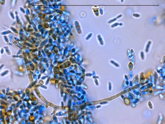
Hortaea werneckii -Another view of a curving septate hyphae surrounded by numerous free conidia.  Annellides can be seen at various points along the hyphae from where the conidia are produced.
(1000X, LPCB, DMD-108)

Hortaea werneckii -The arrow points to an annelide with its developing annelloconidium still attached.  Also seen is a pigmented and septate hyphal element near the top left as well as annelloconidia in various states of maturity.
(1000X, LPCB, DMD-108)

Hortaea werneckii -Another view of a developing annelloconidium attached to the parent annellide.
(1000+10X, LPCB, DMD-108)

Hortaea werneckii -and one more.  Annelloconidium developing in the center of the photo.
(1000+10X, LPCB, DMD-108)
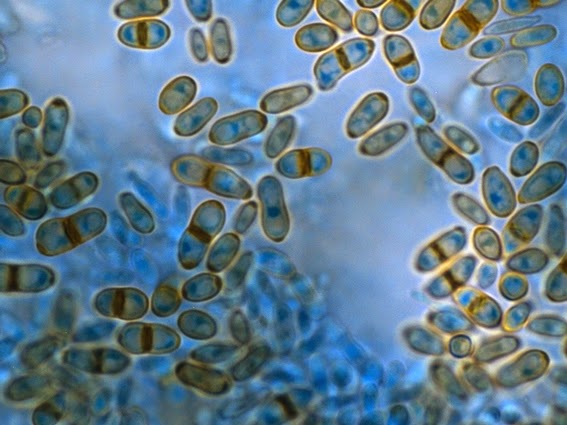
Hortaea werneckii -The primarily two-celled conidia, or more precisely annelloconidia are seen here in various states of pigmentation.  Note that the septum dividing the two cells is darkly stained, as is one end, where the annelloconidium was once attached to its parent annnellide.
(1000+10X, LPCB, DMD-108)

Hortaea werneckii - With aging the annelloconidia may develop into chlamydoconidia-like aggregates (chlamydospores) as seen here in the center of the photograph.
(1000+10X, LPCB, DMD-108)

Hortaea werneckii -The annelloconidia can themselves "bud" and act as annellides, producing annelloconidia as seen here.  
(400X, LPCB, 400X -adhesive tape mount)

Hortaea werneckii -germination of an annelloconidium at top.  Not also the bud developing at the bottom of the two celled annelloconidium.
(1000+10X, LPCB, DMD-108)

Physiology:
-Does not grow at 37˚C
-Nitrate +ve
-Urease +ve
-Halophilic up to 10%

Differentiation:
The annellated zones on Hortaea werneckii are much broader than those of Exophiala species.  Lack of growth at 37˚C also distinguishes the two.


[i] Annellide –A specific conidiogenous cell that produces conidia in succession, each leaving a ring-like collar on the cell wall when released.  ‘Annello’ prefix simply specifies the type of conidiogenous cell or conidia which is produced.

*   *   *

Sunday, 21 October 2012

Scedosporium prolificans


Scedosporium prolificans -Fungus (Previously known as Scedosporium inflatum)

Ecology: Scedosporium prolificans is a cosmopolitan fungus isolated primarily from soils (saprophyte), however its ecosystem and distribution may be more restricted than the other major species, Scedosporium apiospermum. Scedosporium prolificans has been isolated from hospital settings after the environment was disturbed during on-site construction. 
 
Macroscopic Morphology: S.prolificans exhibits moderately rapid spreading growth and will mature within 5 to 7 days at 25oC on sabouraud dextrose agar (SDA or SAB). Young suede-like to downy growth becomes increasingly cottony as it develops an olive-grey to a black colouration. Light coloured mycelial tufts may develop as the colony ages. Reverse is a dark brown to dark grey, almost black in appearance. 

Scedosporium prolificans SAB, 72 hrs,  30oC

Microscopic Morphology: Scedosporium prolificans produces hyaline septate hyphae from which conidiogenous cells are produced along the length. The conidiogenous cells are flask-shaped annellides which have a swollen base (inflated) from which extends an elongated 'neck'. Conidia are produced singly or in small groups at the apex of the annellide. The smooth walled single-celled conidia are hyaline to a pale brown in colour and are oviod to pyridaform in shape. These conidia (annelloconidia) have a truncated base where they were attached to the conidiogenous annellide. The conidia measure 2 - 5 μm to 3 - 13 μm (average 3.4 μm to 5.3 μm) in dimension. 

Note: All photographs which follow were taken using the Leica DMD-108 digital microimaging device. 


Scedosporium prolificans (LPCB, 250X) -First impression of a slide culture.

Scedosporium prolificans -hyphae with conida visible. Conidiogenous cells not all that distinct at this magnification,  (LPCB, 400X)

Scedosporium prolificans -another view of conidia along hyphae
(LPCB, 400X)

Scedosporium prolificans - conidia seen distributed singly and in clusters along the hyphae.  (LPCB, 400X) 

Scedosporium prolificans - At this magnification the conidiogenous cells are becoming more obvious.  Ovoid to pyridaform conidia can be seen attached at the apex of the annellide.  Note 100 micron bar at upper right. (LPCB, 400+10X)

Scedosporium prolificans - Here we can see a conidiogenous cell (annellide) extending from the hyphae at which four annelloconida can be seen at the apex. (LPCB, 1000X)

Scedosporium prolificans - hyphae with annellides bearing ovoid to pyridaform annelloconidia. (LPCB, 1000X)

Scedosporium prolificans - Here we can distinctly see the inflated, vase-like annellide conidiophore (arrows) from which are produced the annelloconidia.  (LPCB, 1000X)


Scedosporium prolificans - Here we can see the elongated 'neck' (1) at the apical end of the annellide and the truncated base or scar at the end of the annelloconidia where it was once attached (2).  The swollen or inflated vase like base is evident just right of (1) where the conidiogenous cell attaches to the hyphae.  (LPCB, 1000X)

Scedosporium prolificans - Here again we can see the elongated 'neck' (1) with an annelloconidia at the apex, the inflated annellide (2) and the annelloconidia, each with a flattened, truncated base of attachment (3).
(LPCB, 1000X)

Scedosporium prolificans - Measurement of annelloconidia - 7.0 μm to 7.4 μm in length, 4.7 μm in diamenter in this field. (LPCB, 1000+10X)

Scedosporium prolificans - small clusters of conidia (annelloconidia) at the apex of annellides (conidiogenous cells) extending from hyphae.
(LPCB, 1000+10X)

Scedosporium prolificans - one last photo showing branched septate hyphae with annelloconidia at apex of annellides.  (LPCB, 1000+10X)


Pathogenicity: Scedosporium prolificans is an emerging fungal pathogen can infect both immunocompetent and immunocompromised individuals. Noted risk factors for scedosporiosis have included malignancy, cystic fibrosis as well as solid organ transplantation. Onychomycosis and mycotic keratitis have been reported in the literature. Inhalation of the fungus has resulted in colonization of the paranasal sinus and the lungs. Penetrating injuries may result in localized infections of the joints resulting in septic arthritis as well as osteomyelitis. Disseminated have been reported in immunosuppressed patients such as those experiencing prolonged neutropenia and post-transplantation therapy. Accurate identification is imperitive as S.prolificans exhibits increased resistance to common antifungal medications and disseminated infections is often fatal even in immunocompetent patients. Asymptomatic colonization as also been reported.

Differentiation: Scedosporium prolificans can be differentiated from Scedosporium apiospermum as the former has a distinctive swollen (inflated) base. This inflated base was the reason this fungus was previously known as Scedosporium inflatum. S.prolificans produces annellides that are flask-shaped, unlike the cylindrical conidiogenous cells of S.apiospermum. S.prolificans is inhibited by media containing cycloheximide.  Finally, S.prolificans has no sexual stage (telemorph) as does Scedosporium apiospermum/Pseudallescheria boydii.


*   *   *










