Showing posts with label trematode. Show all posts
Thursday, 9 January 2014
Fasciola hepatica
Fasciola hepatica (Trematode –
Parasite)
Fasciola hepatica is commonly known as the sheep liver fluke and is a common parasite in
herbivores. With cosmopolitan
distribution, human infections have been reported in many parts of the world. Fasciola
hepatica is most frequently found in countries where sheep raising is
common, such as China, Taiwan, India, Indonesia and other parts of Asia.
Fasciola hepatica is responsible for the disease fascioliasis, also known as
fasciolopsiasis or simply, sheep liver fluke infection. The infection may have first been recognized
as early as 1379 when the effects were noticed between certain water plants and
the sheep that had eaten them. Fascioliasis
is considered a zoonotic disease (passed
from animals to man).
Symptoms: The infection may produce symptoms of biliary
obstruction and cholangitis. Symptoms
may include upper right quadrant pain, fever, chills and jaundice. Symptoms may depend on the worm burden and light
infections may be asymptomatic.
Life Cycle & Morphology:
Worms: The Fasciola
fluke is quite large and may measure as large as 3 cm by 1.5 cm in size. The anterior end of the worm (fluke) has a
distinctive cone shaped projection. The interior organs of the worm appear extensively
branched. The adult worms live in the
bile ducts of the liver and the gallbladder.
Eggs: The eggs (ova) are large (80-150 µm by about
60 -90 µm) and broadly elliptical in appearance. They are operuclated but the operculum is
rather small in relation to the egg and rather inconspicuous. The eggs are unembryonated when passed in the
feces. When passed into water, they
undergo embryonation and subsequently miracidia are hatched (usually in 1 – 2 weeks). Fasciola
hepatica requires an intermediate host for development, which in this case
is a freshwater snail (Lymnaea sp). The miracidia within the snail mature and
emerge as cercariae which then attach to aquatic vegetation (eg. watercress) where they undergo
encystation. Humans are infected by the
ingestion of uncooked aquatic vegetation on which the metacercariae are
encysted. The metacercariae excyst
(hatch) in the duodenum and migrate through the intestinal wall into the
peritoneal cavity. The larvae penetrate the
liver and wander through the parenchyma for up to 9 weeks. The larvae finally enter the bile ducts where
they mature and in about three to four months and begin to produce eggs, which
are ultimately passed out in the feces.
The adult worms may live for up to a year.
Diagnosis:
Diagnosis is made by the
detection of the characteristic eggs in the patient’s faeces. One problem in identification is that the
species Fasciolopsis buski produces
eggs which are almost indistinguishable from those produced by Fasciola hepatica. Life cycles of these two trematodes are very
similar. In some areas of the orient
where these two species overlap, the clinical evaluation of symptoms aids the
diagnosis of these faciolid eggs. The
size of the operculum opening may also assist in diagnosis where the Fasciola hepatica’s operculum is
larger than that of Fasciolopsis buski
(measurements to follow). Putting
pressure on the coverslip of a concentrated faecal specimen with the eraser end
of a pencil may be sufficient to cause the operculum to pop open and better
reveal itself. Molecular methods may
provide a definitive identification.
Fasciola hepatica egg in faecal concentrate. Bile-stained shell and inconspicuous operculum.
Egg measures 151 µm by 75 µm. (DMD-108)
Fasciola hepatica egg with Iron Heamatoxylin stain. (DMD-108)
Fasciola hepatica egg showing the operculum (OP). Measurement reads 28.82 µm. (DMD-108)
* * *
Sunday, 21 November 2010
Schistosoma mansoni & haematobium
Trematodes=flatworms=flukes
Phylum=Platyhelmithes
Family=Schistosomatidae
Primary human species are;
Schistosomes are often referred to as 'blood' trematodes as they differ from other trematodes because they infect humans by penetrating intact skin to gain entry to the circulatory system rather than infection through ingestion. In other words, Schistosomes infect humans through direct larval (cercariae) penetration rather than the ingestion of metacercariae.
Schistosomes are also unique among the flukes in that there are both a male and female organisms.
Schistosoma eggs also lack an operculum which characterizes other fluke eggs.
Their life cycle is as follows;
Symptoms include cercarial dermatitis, acute schistosomiasis (Katayama fever) and related tissue egg deposition. Acute schistosomiasis begins when the adult female begins laying eggs.
In the circualtory system it is believed the organisms either becomes covered with host soluble blood group antigens, lipoproteins, or develops antigens similar to the host's so that it excapes the host's immune response. For this reason, adult worms in the veins evoke little immune response.
Symptoms may vary in intensity but can include malaise, fever, abdominal tenderness or hepatic pain.
Infection with S.mansoni or S. japonicum may cause diarrhea. S. haematobium causes hematuria.
Morphology;
Schistosomiasis should be considered with any patient from endemic areas who has had exposure to untreated water and presents with symptoms previously mentioned. Diagnosis is confirmed with the identification of Schistosome eggs recovered the patient.
All eggs are embryonated when passed
All eggs are easily differentiated by their appearance
S.mansoni & S.japonicum from fecal specimens although on occasion both may be recovered from urine as well.
Treatment;
Praziquantel is the drug of choice in treating schistosomiasis. O & P examinations should be conducted periodically for up to a year post treatement to ensure erradication.
Schistosoma mansoni wallpaper (1024 X 768)
(note lateral 'rose-thorn' spine on egg)
Phylum=Platyhelmithes
Family=Schistosomatidae
Primary human species are;
- Schistosoma mansoni
- Schistosoma haematobium
- Schistosoma japonicum
- (S.mekongi & S.intercalatum encountered less frequently)
Schistosomes are often referred to as 'blood' trematodes as they differ from other trematodes because they infect humans by penetrating intact skin to gain entry to the circulatory system rather than infection through ingestion. In other words, Schistosomes infect humans through direct larval (cercariae) penetration rather than the ingestion of metacercariae.
Schistosomes are also unique among the flukes in that there are both a male and female organisms.
Schistosoma eggs also lack an operculum which characterizes other fluke eggs.
Their life cycle is as follows;
- Eggs in feces or urine are passed into water
- Larvae are liberated and penetrate the intermediate host snail where they further develop.
- Cercariae emerge from the snail while in the water
- Cercariae penetrate the skin of humans in contact with the water
- Larval migration begins through the circulatory system where they may enter alveoli to produce hemoptysis. Organisms mature in the liver before entering specific veins specific to the infecting species. (S.haematobium in veins of the bladder, S.japonicum in veins of small intestine & S.mansoni in veins of the large intestine)
- Eggs are passed to continue the cycle.
Symptoms include cercarial dermatitis, acute schistosomiasis (Katayama fever) and related tissue egg deposition. Acute schistosomiasis begins when the adult female begins laying eggs.
In the circualtory system it is believed the organisms either becomes covered with host soluble blood group antigens, lipoproteins, or develops antigens similar to the host's so that it excapes the host's immune response. For this reason, adult worms in the veins evoke little immune response.
Symptoms may vary in intensity but can include malaise, fever, abdominal tenderness or hepatic pain.
Infection with S.mansoni or S. japonicum may cause diarrhea. S. haematobium causes hematuria.
Morphology;
Schistosomiasis should be considered with any patient from endemic areas who has had exposure to untreated water and presents with symptoms previously mentioned. Diagnosis is confirmed with the identification of Schistosome eggs recovered the patient.
All eggs are embryonated when passed
All eggs are easily differentiated by their appearance
- S.mansoni eggs are large (110-170 µm), oval and have a lateral, 'rose thorn' spine.
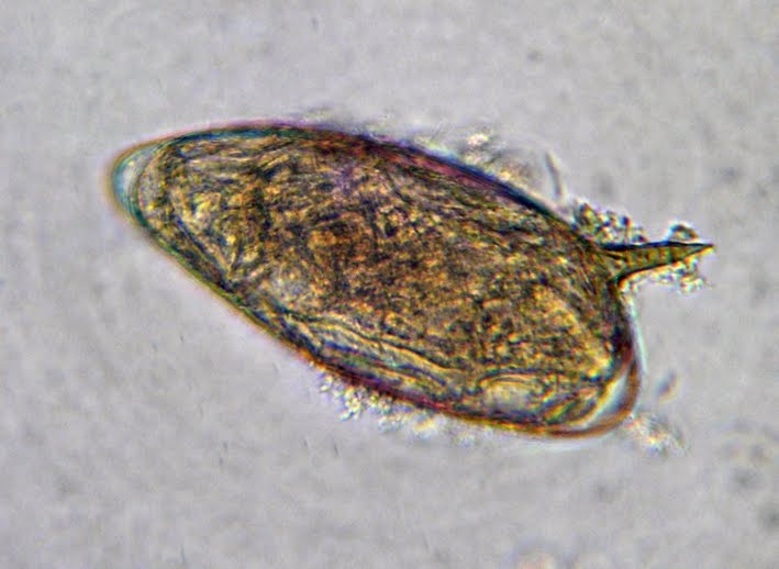
- S.haematobium eggs are large, oval (110-170 µm) and have a terminal (end) spine.
- S.japonicum eggs are smaller (55-90 µm), round and have a 'crooked finger' spine.
S.mansoni & S.japonicum from fecal specimens although on occasion both may be recovered from urine as well.
Treatment;
Praziquantel is the drug of choice in treating schistosomiasis. O & P examinations should be conducted periodically for up to a year post treatement to ensure erradication.
Schistosoma mansoni wallpaper (1024 X 768)(note lateral 'rose-thorn' spine on egg)
I once encountered S.haematobium in the urine of a young Egyptian child however it was prior to my attempts at documenting interesting specimens in photographs. I have never personally seen a S.japonicum.
Update; I recently took some photos of S.haematobium from a preparation obtained from our Pathology department. Unfortunately I don't have much information on this patient's history nor the stain used. However, it does make for a pretty photo!
Schistosoma haematobium
(Note terminal spine)
(Click on photo to enlarge for better viewing)
Schistosoma haematobium (X400)
(Terminal spine at bottom barely visible)
Return Home (Most Recent Posts)
* * *
Update; I recently took some photos of S.haematobium from a preparation obtained from our Pathology department. Unfortunately I don't have much information on this patient's history nor the stain used. However, it does make for a pretty photo!
Schistosoma haematobium(Note terminal spine)
(Click on photo to enlarge for better viewing)
Schistosoma haematobium (X400)(Terminal spine at bottom barely visible)
New -August 2012: Obtained from our pathology department - bladder biopsy with Schistosoma haematobium embedded in tissue. Patient was of middle eastern heritage with relatively recent travel to that region but I have no further information. Schistosoma egg is in a deteriorated state in these tissue section photos, leaving not much more than the outline with little internal structure remaining.
Deteriorated S.haematobium embedded within bladder tissue (X500 Nikon: Giemsa?)
Two deteriorated S.haematobium eggs embedded in bladder tissue. (X500 Nikon: Giemsa?)
New -Added November 08, 2013
Fresh urine sample from patient from Africa. Total urine sample was centrifuged to concentrate and re-suspended sediment in a smaller volume. Wet preparation was examined under the light microscope.
Schistosoma haematobium -unstained concentrate
(400+10X, DMD-108)
Schistosoma haematobium -unstained concentrate (note terminal spine)
(400X, DMD-108)
Schistosoma haematobium -unstained concentrate
Still fresh, the parasite was still alive with continuous movement and cytoplasmic streaming within the organism. (400X, DMD-108)
Return Home (Most Recent Posts)
* * *
Subscribe to:
Posts (Atom)