Friday, 24 February 2012

Absidia corymbifera (Lichtheimia corymbifera complex)


Mycocladus corymbifer (formerly Absidia corymbifera)
Lichtheimia corybifera complex (formerly Absidia corymbifera) Fungus
(Names seem to change monthly!!! - your choice!) 
Absidia corymbifera;
I’m going to refer to this organism throughout this post by its former name, “Absidia” corymbifera as most are still unfamiliar with the change in nomenclature.
Pathogenicity:
Although uncommon, Absidia species have been implicated as an agent of human (sub)cutaneous mycosis as well as opportunistic pulmonary infections. As with other fungi previously considered to be relatively benign, they are now being isolated with greater frequency particularly in immunocompromised patients (AIDs, transplant patients, neutropenic patients, etc.) Absidia corymbifera is considered to be the only species of Absidia that is recognized as a human pathogen. Absidia corymbifera occasionally has been known to cause mycotic abortions in cows.
Ecology:
Absidia species are ubiquitous in nature and can be isolated from soils and decaying vegetation. As such they may be found as contaminants in cultures. Care must be taken in determining their significance when isolated.
Macroscopic Appearance:
Colonies grow rapidly, maturing in 3 – 4 days. They are white to a greyish-brown in colour with a woolly texture also described as resembling ‘cotton candy’. Reverse is uncoloured and non-descript. Absidia species are inhibited by cycloheximide and therefore will not grow on media such as mycosel. Maximum growth temperature for Absidia corymbifera is between 45 – 52oC. Absidia corymbifera is thermophillic in that it grows more rapidly at 37oC than at 25oC accounting for its human pathogenicity (body temperature). Most other Absidia species fail to grow at 37oC.
Absidia corymbifera on SAB media after 4 days incubation at 30oC
(Reverse not shown - rather nondescript colourless)
Microscopic Morphology:
Absidia’s hyphae are aseptate (rare septa) and relatively wide at 6 – 15 µm in diameter. Absidia species resembles Rhizopus species however the distinguishing feature is that the sporangiophores of Absidia arise from locations on the stolon that lie between the rhizoids and not from them. In other words, there is no rhizoid directly below the sporaniophore as would be observed in Rhizopus species.
Sporangiophores arise alone or in groups from the aerial hyphae and are rather long (up to 450 µm). Sporangiophores branch and widen apically where they form a conical apophysis immediately below the columella. Sporangia are about 20 – 90 µm in diameter are spherical to pyriform in shape. When mature the sporangial wall dissolves releasing round to oval (3 – 5 µm) sporangiospores leaving a small collarette where the apophysis and sporangium had met. Zygospores have been described.
Note: Apophysomyces elegans resembles Absidia corymbifera but produces white colonies and fails to sporulate on routine clinical laboratory media.
Photos below taken with the Leica DMD-108 Microscope.
Young slide culture Absidia corymbifera X100 LPCB
(Click on any photo to enlarge for better viewing)
Absidia corymbifera - Structures seen; (LPCB X100)
  1. Rhizoid
  2. Sporangiophore arises from stolon between rhizoids and not directly from a rhizoid though this appears to be more of a branch in this photo. Rhizoids were a bit of a challenge to find on this particular organism.
  3. Sporangiophore
  4. Sporangium
Ditto (LPCB X100)
Rhizoids were rather difficult to detect. Adhesive tape preparations failed to capture any. I suspect they were so well anchored to the media that the sporangiophores broke off from the stolon rather than pulling the stolon and rhizoids out with it. This photo was taken from a slide culture preparation where some of the agar media surrounding the rhizoid was removed along with the stolon and sporangiophore.
Numerous sporangia with loose sporangiospores are pictured. Notice that the sporangiophore widens apically where it becomes the columella and supports the sporangium. (LPCB X400)
Ditto
Absidia corymbifera sporangium filled with sporangiospores. (LPCB adhesive tape preparation X400 + additional 1% digital magnification)
More views of the same. Apophysis structure can be seen in dissolving sporangium on left. (LPCB X400)
Ditto
Sporangium dissolvingé breaking up releasing Sporangiospores
(LPCB X400)
Apophysis with remains of Collarette and a few remaining Sporangiospores.
More views of same - Stolon with branching sporangiophores bearing Sporangia in various stages of releasing Sporangiospores. Apophysis can be visualized on Sporangiophore at upper left. (LPCB X 400)
Dissolving sporangium releasing numerous sporangiospores with remaining apophysis on apical end of sporangiophore. (LPCB X400)
Wednesday, 22 February 2012

Strongyloides stercoralis Revisited (Sputum)

Parasite (Nematode)

While I already had a 2010 post on Strongyloides stercoralis , I decided to add a separate posting for this case as it differs in that the worm was not obtained from a fecal sample but rather from a sputum sample. It is precisely these twists that make microbiology both fun and challenging.

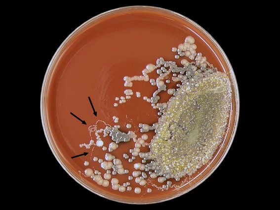
An astute colleague was examining a routine sputum culture for bacterial pathogens on this patient when he picked up on a clue that many might have missed. Small looping 'threads' (for lack of a better description) of bacteria which extended from the primary growth (see photo below). He immediately realized that this might be evidence of the presence of a worm. A viable (live) worm if present in the sputum sample would be inoculated onto the agar media along with respiratory bacteria. They can crawl just as an earthworm can crawl on a wet sidewalk. As they migrate around the plate they draw along bacteria that have adhered to their sides and deposit these along the way providing evidence of their travels. With continued incubation the individual deposited bacteria grow up into colonies outlining the worms journey.

Above (Arrows); A chocolate agar plate inoculated with a sputum sample showing curious looping trails of bacteria which were deposited by a worm migrating about the plate during incubation. Having noticed this curious evidence, the following morning the plate was examined under a plate microscope however the worm responsible for making this trails was not located.

Strongyloides stercoralis can migrate out of the gastrointestinal tract and pass through the lungs. The worm can be coughed out and/or re-swallowed to reinfect the patient. The attending physician had noted that this patient had produced "coffee ground" emesis (hematemesis). This is partially digested blood tinged sputum coughed up, or perhaps swallowed, then coughed up. It has the apperance of wet coffee grounds, hence the description. The sputum sample received by our lab no longer showed this coffee ground appearance. Our patient was an elderly gentleman with several other underlying medical conditions which will not be noted here.

Two Strongyloides stercoralis worms seen in this gram stain of sputum - one curled and one straight directly above. White blood cells dot the background.
Note micron bar in upper right corner. (DMD-108 Microscope X100)

Same worms as above (Gram Stain X1000)

Another Strongyloides stercoralis worm (note striated texture of body)
(Gram Stain of Sputum - DMD-108 Microscope: X1000)

Anterior end of worm with buccal cavity visible.
Compare size to gram negative bacilli bacteria present.
(Gram Stain of Sputum -DMD-108 Microscope X1000)

Strongyloides stercoralis in Iron Hematolxyin Stained Sputum Isolate.
(DMD-108 Microscope X1000)

Two Strongyloides stercoralis worms seen in this Iron Hematoxylin stained preparation of the sputum sample received. Numerous white blood cells present.
(DMD-108 Microscope X100)

Having requested additional specimen samples (and stool sample for interest sake), Strongyloides stercoralis eggs containing developing larvae were also found.
Above; Both a worm and egg are present with the egg trailing some incidental material attached to one end. (Concentrate of sputum sample DMD-108 Microscope X100)

Same egg as above with extraneous material trailing off to lower right
(DMD-108 Microscope X400)

Another Strongyloides stercoralis egg containing developing larvae
(Concentrate - DMD-108 Microscope X400)

Strongyloides stercoralis worm in sputum.
(Concentrate - DMD-108 X400)

A stool sample was received by the lab however diagnosis had already been made starting by those rather inconspicuous tracks left by the worm on bacterial agar media.

For further information on clinical symptoms, diagnosis etc, please refer to my previous post located here; Strongyloides stercoralis
Return to top (Most Recent Posts)

Sunday, 5 February 2012

Microsporum canis

Microsporum canis; (Fungus)

Pathogenicity;

Microsporum canis is a cosmopolitan zoophilic dermatophyte usually acquired from infected dogs (hence canis) or cats. Most prevalent in children, it has been implicated in infections of the scalp and skin and occasionally nails. A scalp infection could be visualized by examining plucked hairs under a Wood’s (UV/black light) lamp for fluorescence.

Colony Morphology;

Surface growth has been described as downy to woolly, to fluffy, hairy and silky. Typically it exhibits a light yellowish pigment at the periphery and growth shows closely spaced radial grooves. The reverse is pale tan to yellowish (yellowish-orange –media dependent) which tends to turn brownish as it ages.

Microsporum canis on SAB at 5 days at 30oC

Microscopic Morphology;

Microsporum canis has septate hyphae that produce numerous macroconidia. The macroconidia are rather long (10-25 X 35-110 µm), spindle or fusoid in shape and are thick walled with a echinulte (rough) texture. The ends typically taper to a knob-like end that may be somewhat recurved at the apex. The macroconidia usually have six or more compartments when mature and few smooth walled club shaped macroconidia may be observed along the hyphae. Smooth walled, club shaped microconidia are infrequently seen forming along the length of the hyphae.

(all photos taken with DMD-108 digital microscope except where noted)

Microsporum canis growing at edge of cover slip from a slide culture preparation (LPCB X100)

(Click on any photo to enlarge for better viewing)

Ditto

Microsporum canis (LPCB X100) Nikon

Microsporum canis (LPCB X250)

Microsporum canis (LPCB Magnification not noted)

Microsporum canis (LPCB X400) -Nikon

Microsporum canis - Macro & microconidia (LPCB X400)

(note 100 µm bar at upper right of photo)

Microsporum canis compartmentalized macroconidia showing rough surface with knob-like end (LPCB X1000 Adhesive Tape Preparation) -Nikon

Ditto (note curved ends)

Microsporum canis - 10% KOH* preparation (X400)

Microsporum canis - 10% KOH* preparation (X400)

(Inset; 2 macroconidia)

Misc;

Microsporum canis needs no special growth factors or cultural requirements. I found that on the relatively nutritious Sabouraud Dextose agar, my isolate became sterile on repeated subcultures. Macroconida were produce when grown on Corn meal agar. The macroconidia are produced as a survival mechanism and are induced when conditions are not as rich or favourable for growth.

Microsporum canis can be inoculated onto sterile (autoclaved) polished rice grains where they produce a yellow pigment.

A hair perforation test (positive) can be performed in vitro. As previously stated, hairs infected with Microsporum canis will fluoresce under a Wood’s UV lamp.

* * *

*10% KOH - Potassium Hydroxide, kills and clears the preparation.

Home: Return to most recent posts.

Saturday, 4 February 2012

Aspergillus flavus

Aspergillus flavus (fungus)

Ecology & Pathogenicity;

Found worldwide. Ubiquitous in nature and is the second most common cause of invasive aspergillosis next to Aspergillus fumigatus. Has been implicated in pulmonary, systemic, sinus, ear and other infections. Most widely reported food-borne fungus and can be found colonizing decaying vegetation, crops and seeds. Has also been implicated as both an insect and animal pathogen. (For a slightly more expanded description of pathogenicity, see the intro to the Aspergillus fumigatus post.)

Macroscopic Colony Morphology;

Very rapid rate of growth, maturing in about three days. Surface is greenish-yellow to olive and may have a white border. Texture is often floccose, especially near the center and overall can be velvety to woolly. Unremarkable cream to tan to yellowish reverse on Sabouraud Dextrose media.

Aspergillus flavus on SAB media at 72 hour at 30oC

(note greenish-yellow colour with white edge)

(click on any photo to enlarge for better viewing)

Microscopic Morphology;

Septate hyphae with rather long conidiophores (~400-800 X 8-17 µm) which have a rather rough texture or even spiny, especially just below the vesicle. Vesicles are spherical to elongate and about 20 – 45 µm wide. Aspergillus flavus can be variable in seriation with most strains being about 20% biseriate however some strains can be almost entirely uniseriate. Metulae (8-10 X 5-7 µm) cover three quarters to the entire surface of the vesicle from which the phialides (7-12 X 3-4 µm) form. The metulae support the phialides and together form the biseriate structure. Conidia are globose to ellipsoidal (3-6 µm) with smooth to finely roughened walls.

(Photos below are of a ~30hr old slide culture preparation taken with the DMD-108 digital microscope - except where noted)

Aspergillus flavus mycelium &, conidiophores (LPCB) (X250)

Tangled web of the Aspergillus flavus mycelium as above (X250)

Aspergillus flavus conidiophore bearing vesicle (X1000)

look for the biseriate structure (metulae & phailide) on this and the following photos

Aspergillus flavus vesicle bearing metulae & phialides from which forms the conidia

Aspergillus flavus (LPCB X1000)

Look for the Biseriate structure - Rough surface of conidiophore at its apex (where it meets the vesicle ) is evident in this photo. This roughness is a diagnostic feature of Aspergillus flavus.

Aspergillus flavus (LPCB X1000) Biseriate structure may be easier seen in this photo.

Aspergillus flavus (LPCB X1000)

(Too many photos, but I just like them!)

Aspergillus flavus (LPCB X1000 + 10X digital magnification from DMD-108 Scope)

Though difficult to see, the slighly rough wall can be visualized on several of the conidia, particularly in the lower left of the photo. The conidiophore also has this rough or gently spiked texture particularly at the apex (where it meets the vesicle)

Also note that phialides radiate from vesicle in all directions as opposed to A.fumigatus where they tend found on the upper 2/3rds of the vesicle and extend parallel to the conidiophore.

Aspergillus flavus (LPCB X1000)

(again, enlarge photo and look for the rough texture of the conidia's surface)

Aspergillus flavus - more typical appearance with phialides radiating from vesicle in all directions. This photo taken from adhesive tape preparation of 72 hr fully mature culture. This technique was quite disruptive, dispersing conidia throughout the preparation.

(LPCB X400 -Nikon)

Mycotoxins;

Many Aspergillus flavus isolates are capable of producing aflatoxins, very potent carcinogens. (Aflatoxin B1, cyclopiazonic acid, 3-nitroproprionic acid.)

Intended as Aspergillus flavus computer screen 'Wallpaper' (1024X768 when posted)

Home: Return to Most Recent Posts

Friday, 20 January 2012

Aspergillus fumigatus

Aspergillus fumigatus (Fungus)

Aspergillus species (in general);

There are about 180 species of Aspergillus, 20 of which are recognized as being human pathogens. Aspergillus fumigatus is the species most commonly implicated, followed by Aspergillus flavus and Aspergillus niger.

Pathogenicity;

Most commonly, Aspergillus infections in humans are pulmonary (ie. involve the respiratory system) however they can disseminate to produce deep seated infections particularly in immunocompromised individuals such as transplant and neutropenic patients. Aspergillus may also cause allergic aspergillosis and fungal sinusitis. Aspergillus is also capable of causing illness in other species such as birds and cattle.

Ecology;

Aspergillus fumigatus is cosmopolitan & ubiquitous. Primarily a saprobic fungus of soils which can be found living on substrates such as plants, compost, seeds and wood chips, etc. Hospital renovations have been implicated in the dissemination of Aspergillus fumigatus which can be detrimental to the patient and a nuisance contaminant for the laboratory. Careful consideration must be given before dismissing Aspergillus fumigatus as a laboratory culture contaminant.

Physiology;

A.fumigatus is rapidly growing fungus which matures in about 3 days. Aspergillus fumigatus is thermophillic, with good growth to 45oC and often up to 50oC. This ability can be used to differentiate it from other species.

Colony Morphology;

The surface growth is velvety, downy or powdery, showing various shades of green, most commonly a blue-green to a grey-green with a narrow white border. The colour typically darkens with age. The reverse is white to tan to pale yellowish. Colouration or shade can be dependent on the media on which the fungus is cultured upon.

Aspergillus fumigatus on SAB Agar after 5 days incubation at 30oC (Surface)
(Click on any photo to enlarge for better viewing)

Microscopic Morphology;

Hyphae are septate with smooth walled conidiophores (usually less than 300 µm in length and 5-10 µm wide). Vesicles are subclavate in shape, roughly 20-30 µm in width. Conidiogenous cells (phialides) are flask shaped, uniseriate, compact (closely spaced), usually forming on the upper two-thirds of the vesicle and mature parallel to the axis of the conidiophore (columnar formation). Young conidial heads may radiate. Conidia are verrucose (smooth or slightly rough), (sub)spherical and about 2-3.5 µm in diameter and develop in chains.

Photos below were taken with the DMD-108 digital microscope. Magnifications are as noted. In my opinion, the uniseriate phialides (or biseriate in other species) are best visualized and determined under oil. The DMD-108 microscope has an addition optional digital magnification factor of X10 which can be utilized for even greater enlargement of features. Both adhesive tape and slide culture preparations were made and stained with Lactophenol Cotton Blue (LPCB). The formed chains of conidia were easily disrupted even with the slide culture technique. A little disappointing however the photos below were the result.

(Most photos taken with the DMD-108 digital scope, Nikon where noted.)

Structures described in test above shown for clarity (X1000 oil immersion)
(Click on photo to enlarge for better viewing)


Aspergillus fumigatus (LPCB - Adhesive tape preparation) (X250)

Aspergillus fumigatus (LPCB - Adhesive tape preparation) (X400)
(Phialides generally on upper 2/3 of vesicle and more or less parallel to the conidiophore axis)

Ditto (X400)
(Structure size may vary between photos under same stated magnification as a result of how the image was cropped for publishing)

(X400) Nikon

Aspergillus fumigatus conidiophores (LPCB) (X400)

Aspergillus fumigatus conidiophores & free conidia (LPCB) (X400)

Ditto

Aspergillus fumigatus conidiophore & conidia (LPCB) (X1000)

Ditto

Aspergillus fumigatus (LPCB) (X1000)

Mycotoxins;

Aspergillus fumigatus produces a variety of mycotoxins, the major ones being Gliotoxin, verrucologen, fumitremorgin A & B.

Intended as computer 'wallpaper' (1024 X 768)










