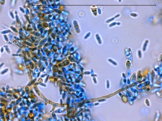
Microscopic Morphology: Cladosporium produces erect, dark,
septate hyphae. Conidiophores are also
darkly pigmented, may be septate and show tree-like branching. Fragile chains of dematiaceous blastoconidia
are produced and exhibit a dark hila or scar at their point of attachment to
the conidiophore or other conidia. The
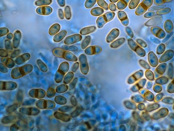
1-4 celled conidia are round to oval (3 -6 µm X 4 - 12 µm) and may be
smooth-walled to verrucose in surface texture.
Cells on the conidiophore which bear the chains of conidia are sometimes
septate and appear in the shape of a ‘shield’.
These cells are also conidia but are referred to as shield cells. Chains of conidia easily disarticulate (break
up) and were frustratingly difficult to document using both adhesive tape andslide culture techniques.Saturday, 14 June 2014
Cladosporium species (Revisitied)
Cladosporium species Revisited (Kind
Of) -A Black Mould -Hyphomycetes
This post is a bit of a revisitation to the fungus Cladosporium as I previously included
the genera in a previous post on Cladophialophoraspecies. You can compare the two more
thoroughly by checking out my previous post by clicking here.
Ecology: Cladosporium species
are cosmopolitan saprophytic fungi found in soil, on plant debris and leaf
surfaces.
Pathogenicity: Cladosporium
species are generally considered to be non-pathogenic although they may be
considered as possible opportunists in the severely debilitated host. Potentially pathogenic species previously
included under the Cladosporium
genera have recently been reassigned to the genera Cladophialophora (eg.
C.bantianum, C.carrionii). Cladosporium may be encountered in the
laboratory as a culture contaminant and must be distinguish from the pathogenic
Cladophialophora.
Macroscopic Morphology: The rate of growth is dependent on the
particular Cladosporium species and
can vary from slow to moderately rapid.
The isolate discussed in this post expanded in size rather slowly though
it matured to produce copious amounts of conidia rather quickly (~7 days). The colony was velvety to suede-like in
texture. Other sources describe Cladosporium’s texture as ranging from
powdery to woolly. The colony may become
slightly heaped and develop gentle folds as it ages. Colour ranges from greyish-green to
olivaceous-green to brownish-black. The
reverse is a dark brown to black in colour.
Cladosporium species on SAB, 15 Days at 30˚C (Nikon)
Cladosporium species on SAB, 25 Days at 30˚C (Nikon)
Microscopic Morphology: Cladosporium produces erect, dark,
septate hyphae. Conidiophores are also
darkly pigmented, may be septate and show tree-like branching. Fragile chains of dematiaceous blastoconidia
are produced and exhibit a dark hila or scar at their point of attachment to
the conidiophore or other conidia. The
1-4 celled conidia are round to oval (3 -6 µm X 4 - 12 µm) and may be
smooth-walled to verrucose in surface texture.
Cells on the conidiophore which bear the chains of conidia are sometimes
septate and appear in the shape of a ‘shield’.
These cells are also conidia but are referred to as shield cells. Chains of conidia easily disarticulate (break
up) and were frustratingly difficult to document using both adhesive tape andslide culture techniques.
Cladosporium species are not thermotollerant and some
species may not grow at 37˚C.
Cladosporium species produce darkly pigmented, septate, branching hyphae,
(LPCB, DMD-108, 400+10X)
Cladosporium species - Conidiophores are also darkly pigmented, may be septate and also show tree-like branching. (LPCB, DMD-108, 400+10X)
Cladosporium species - The Lactophenol cotton blue stain has taken more deeply in this preparation. Septate hyphae are clearly visible. (LPCB, DMD-108, 400X)
Cladosporium species -1-4 celled conidia are round to oval (3- 6 µm X 4-12 µm) and may be smooth-walled to verrucose in surface texture.
(LPCB, DMD-108, 1000X)
Cladosporium species -the structures are clearly evident here: septate hyphae, darkly pigmented conidiophores and oval conidia in chains.
(LPCB, DMD-108, 1000X)
Cladosporium species -as above. Younger conidiophores & conidia stain more intensely with the LPCB while the mature structures have developed the pigmentation.
(LPCB, DMD-108, 1000X)
Cladosporium species -the structures easily dis-articulate (break up) and it was difficult to keep them intact regardless of using the adhesive tape technique or slide culture technique.
Short chains of oval, pinmented, conidia are seen.
(LPCB, DMD-108, 1000X)
Cladosporium species - a closer look at the conidiophores bearing round to oval pigmented conidia.
(LPCB, DMD-108, 1000+10X)
Cladosporium species -a single conidia (conidiophore?) seen at the end of a septate hyphae.
(LPCB, DMD-108, 1000X)
Cladosporium species - a structure visible in some of the previous photos is more clearly seen in this picture. Here you can clearly see what is referred to as a "shield cell", because of its resemblance to a warrior's shield. These are conidiophores as you can clearly see chains of conidia extending from them. (LPCB, DMD-108, 1000+10X)
Cladosporium species - `Shield` cell conidiophore and conidia
(LPCB, DMD-108, 1000+10X)
Cladosporium species - another view, as above.
(LPCB, DMD-108, 1000+10X)
Cladosporium species - `Shield`cells and chains of conidia
(LPCB. DMD-108, 1000X)
Cladosporium species - Septate hyphae with conidiophore bearing conidia at its apex. Insert is simply a change of focus. (LPCB, DMD-108, 1000X)
Cladosporium species - okay, one final photo, just for the heck of it!
(LPCB, DMD-108, 1000+10X)
* * *
Friday, 18 April 2014
Chilomastix mesnili
Chilomastix mesnili (Flagellate)
Intestinal Parasite
Note: I'm always hesitant in adding posts of organisms where the photos don't clearly show the features or structures described. Such is the case here -fine detail is not evident, but here are the photos for whatever they are worth.
Geographic &
Human Habitats:
In nature, Chilomastix
mesnili is considered to be cosmopolitan and therefore has worldwide
distribution, found in both warm & temperate climates. In humans, it can be found primarily in the
cecal region of the large intestine, but may also occur in the small
intestine. There it feeds on intestinal
bacteria and debris.
Pathogenicity:
C.mesnili is
considered to be non-pathogenic and treatment is not warranted. Its presence in stool specimens does indicate
that the host has acquired the parasite through the ingestion of its cysts and
therefore has come in contact with contaminated material. Further examination for other more serious
parasites might be advised. Personal and
public hygiene are key in the prevention this parasite.
Morphology:
Chilomastix mesnili
has both trophozoites and cyst forms.
Diagnosis is made by detecting one or both of these forms in fecal
specimens.
Trophozoites: ‘Trophs’ are
elongate, tapering towards the posterior end.
They can measure 6 µm to 24 µm in length but the more common range is 10
µm to 15 µm. In a freshly obtained
stool sample, the trophozoites exhibit a ‘stiff’, rotary motion. The nucleus is generally not recognizable in
unstained samples from lack of contrast.
There are three anterior flagella and a groove running in a spiral
manner along the length of the organism.
In stained preparations, the single nucleus can be seen, located near
the origin of the anterior flagella. The
cytostome, or oral depression, is bordered by fibrils extending one-third to
one-half down the length of the organism.
The most prominent cytostomal fibril curves posteriorly around the cytostome
and resembles a ‘shepherd’s crook’. The
flagella stain poorly, if at all. The
nucleus is found at the anterior end of the organism. Peripheral nuclear chromatin may be present
in the form of granules or irregularly distributed as plaques against the
nuclear membrane. A small, central or
eccentric karyosome may at times be seen.
Chilomastix mesnili -trophozoite seen here with a large nucleus in the anterior end with the flagellate tapering posteriorly. The karyosome is seen to be eccentric. Some inclusions are visible within the cytoplasm.
(Iron-Hematoxylin Stain, 1000X, Nikon)
Chilomastix mesnili -troph as described in the previous photo. Karyosome is not obvious.
(Iron-hematoxylin, 1000X, Nikon)
Chilomastix mesnili -The previous two photos were cropped to better isolate the organism. This un-cropped photo better shows what the Chilomastix mesnili troph would appear like under the light microscope at 1000X magnification when analyzing a stool specimen. Parasitology is a science, skill and art combined, making it one of the more challenging disciplines in clinical microbiology to master.
(Iron-hematoxylin, 1000X, Nikon)
Cysts: The cyst formed by C.mesnili is typically described as
lemon or pear shaped, with a prominent knob-like protuberance at the anterior
end. (Appearance always depends on the
orientation of the organism when fixed and stained.) This shape is characteristic of Chilomastix mesnili alone and does not
appear in any other intestinal protozoa.
Cysts can range from 6 µm to 10 µm in size but generally average 7 µm to
9 µm in length, with the width being slightly less. The cyst contains a relatively large nucleus
and the chromatin may be condensed to form a large karyosome. Curved cytostomal fibrils are usually quite
prominent and frequently may give the appearance of an open safety pin,
alongside the cytostome.
Chilomastix mesnili -cyst. Note the slightly distorted shape giving the cyst a lemon or pear-like appearance. A rather large nucleus is seen with abundant peripheral chromatin.
(Iron-hematoxylin, 1000X, Nikon)
Chilomastix mesnili -as above. The skill is to find and identify parasites when often hidden or masked by fecal debris present after processing.
(Iron-hematoxylin, 1000X, DMD-108)
Chilomastix mesnili - due to the orientation, it is difficult to tell in this photo. The nucleus appears at one end suggesting a trophozoite, however it may also be a cyst. Actual size measurement was not recorded for this photo.
(Iron-hematoxylin, 1000X, Nikon)
Chilomastix mesnili -cyst
(Iron-hematoxylin, 1000X, DMD-108)
Chilomastix mesnili -cyst (middle of photo) -other structures are artifacts.
(Iron-hematoxylin, 1000X, Nikon)
Chilomastix mesnili - Chilomastix troph (C.m.) seen above and to the left of an Entamoeba coli cyst (E.c.) -for size comparison.
(Iron-hematoxylin, 1000X, DMD-108)
Chilomastix mesnili -cyst (Cm) seen. A Blastocystis hominis (Bch) cell seen at top of photo. Cell seen near middle of photo shows to few characteristics for definitive identification but may be another Chilomastix organism.
(Iron-hematoxylin, 1000X, DMD-108)
Chilomastix mesnili - cyst in an unstained concentrate. Can you find it without an arrow pointing to it?
(Fecal concentrate, 400X, Nikon)
Chilomastix mesnili - trophozoite in an unstained fecal concentrate.
(Fecal concentrate, 400X, Nikon)
Again, the morphology of Chilomastix mesnili makes it
rather easy to identify in well stained preparations. However, both trophs and cysts may stain
lightly making them difficult to distinguish when mixed in with fecal material. It must be distinguished from other small
parasites that may be present such as Enteromonas
hominis and Retortamonas intestinalis.
* * *
Monday, 31 March 2014
Hortaea werneckii
Hortaea werneckii -Mould (Phaeoannellomyces werneckii)
Ecology:
Hortaea werneckii is a saprophytic
fungus which can be found in tropical and sub-tropical soils, compost, and
decaying wood. This fungus is also halophilic
as it has been isolated from salted foods, saline waters and natural saltpans,
growing in salt concentrations of up to 10%.
Pathogenicity:
H.werneckii is
the etiological agent of superficial skin infections known as tinea nigra. This infection usually manifests itself as
brown to black macules which are most often found on the palms of the
hands. The appearance may initially resemble
that of malignant melanoma.
Macroscopic
Morphology:
Hortaea werneckii
is a rather slow growing fungus which initially appears as shiny black, slimy
or mucoid yeast. As the colony matures,
development of aerial hyphae may give the colony a velvety texture. The reverse also has a rather non-nondescript
brown-black colour.
Hortaea werneckii SAB 16 days at 30˚C (Nikon)
Reverse: (not shown) appears much like the surface, perhaps slightly more olivaceous in colour.
Right: single colony incubated for approximately one month shows a slight velvety texture.
Aerial mycelia develop and acquire the olivaceous black
colour as they age. Septate hyphae may
be rather wide, reaching up to 6 µm in width.
Intercalary or lateral conidiogenous (annellide[i])
cells develop along the hyphae which produce the conidia (annelloconidia). When released the annelloconidia show a
prominent annellated ring, 1 -2 µm wide where once attached.
Annelloconidia are also hyaline (clear) becoming
translucent olivaceous brown-black in maturity.
They are smooth-walled and have a broad ellipsoidal appearance (7.0 –
9.5 µm X 3.5 – 4.5 µm). Annelloconidia
are 1 to 2 celled and the internal cell wall or septum is usually deeply
pigmented. With aging the annelloconidia
may develop into chlamydoconidia-like aggregates (chlamydospores).
Hortaea werneckii - At low magnification there is not much to see other that a sea of cells (conidia)
(KOH, 250X, DMD-108)
Hortaea werneckii -brown or olivaceous pigmented conidia are visible
(KOH, 400X, DMD-108)
Hortaea werneckii - two celled conidia can now be distinguished.
(KOH, 400+10X, DMD-108)
Hortaea werneckii -Pigmented, septate hyphae with lateral annellides.
(LPCB, 1000X, DMD-108)
Hortaea werneckii -Here we can see the broadly ellipsoidal, two-celled annelloconidia. Mature conidia have the brown pigmentation while younger cells stain more intensely with the Lactophenol Cotton Blue (LPCB). One end of the annelloconidia usually stains darker indicating the location of the annellated ring or the point where it was previously attached to the conidogenous annellide.
(1000X, LPCB, DMD-108)
Hortaea werneckii -Another view of a curving septate hyphae surrounded by numerous free conidia. Annellides can be seen at various points along the hyphae from where the conidia are produced.
(1000X, LPCB, DMD-108)
Hortaea werneckii -The arrow points to an annelide with its developing annelloconidium still attached. Also seen is a pigmented and septate hyphal element near the top left as well as annelloconidia in various states of maturity.
(1000X, LPCB, DMD-108)
Hortaea werneckii -Another view of a developing annelloconidium attached to the parent annellide.
(1000+10X, LPCB, DMD-108)
Hortaea werneckii -and one more. Annelloconidium developing in the center of the photo.
(1000+10X, LPCB, DMD-108)
Hortaea werneckii -The primarily two-celled conidia, or more precisely annelloconidia are seen here in various states of pigmentation. Note that the septum dividing the two cells is darkly stained, as is one end, where the annelloconidium was once attached to its parent annnellide.
(1000+10X, LPCB, DMD-108)
Hortaea werneckii - With aging the annelloconidia
may develop into chlamydoconidia-like aggregates (chlamydospores) as seen here in the center of the photograph.
(1000+10X, LPCB, DMD-108)
Hortaea werneckii -The annelloconidia can themselves "bud" and act as annellides, producing annelloconidia as seen here.
(400X, LPCB, 400X -adhesive tape mount)
Hortaea werneckii -germination of an annelloconidium at top. Not also the bud developing at the bottom of the two celled annelloconidium.
(1000+10X, LPCB, DMD-108)
Physiology:
-Does not grow at 37˚C
-Nitrate +ve
-Urease +ve
-Halophilic up to 10%
Differentiation:
The annellated zones on Hortaea werneckii are much broader than those of Exophiala species. Lack of growth at 37˚C also distinguishes the
two.
[i]
Annellide –A specific conidiogenous cell that produces conidia in succession,
each leaving a ring-like collar on the cell wall when released. ‘Annello’ prefix simply specifies the type of
conidiogenous cell or conidia which is produced.
* * *
Subscribe to:
Posts (Atom)


.jpg)


.jpg)
.jpg)