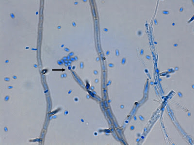
March 29th, 2014 Update: My thanks to Eduardo from Chile who contacted me suggesting that this black mould may be a Tritirachium species, possibly dependens. Checking one of the reference books that I have listed in the left sidebar 'Pictorial Atlas of Soil & Seed Fungi' I see that Eduardo may be correct. Thank you!
Saturday, 29 December 2012
UnIdentified Black Mould
Unidentified Environmental Black
Mould No.1
March 29th, 2014 Update: My thanks to Eduardo from Chile who contacted me suggesting that this black mould may be a Tritirachium species, possibly dependens. Checking one of the reference books that I have listed in the left sidebar 'Pictorial Atlas of Soil & Seed Fungi' I see that Eduardo may be correct. Thank you!
* * *
As the title of this Blog Site suggests, Microbiology
should be Fun! The pursuit of knowledge
should be as enjoyable as discovery - the journey as much fun as the
destination…especially if one has yet to arrive.
Presented in this post is an environmental isolate whose
identity has of yet eluded me. (No.1 as I'm sure I'll have more) As my resources
are primarily medical, environmental fungi which have little or no medical importance
are poorly, if at all documented. Regardless,
one quick look at a hastily prepared adhesive-tape mount convinced me to have a
closer look and to share a few photos. Perhaps
a reader of this post might offer a clue or steer me in the right direction so
as to give this dark, spiky fellow a proper name.
Ecology: As mentioned, this isolate was an
environmental contaminant and its distribution and preferred habitat remains
unknown.
Macroscopic
Morphology: Colonies are slow
growing, reaching one to two centimeters in diameter in 10 – 14 days at 30oC. Growth has yet to be tested at 37oC
or 40 – 45oC. Growth starts
off white but quickly develops a dark olivacious to smoky-grey to black appearance. A small white outer margin may remain. Overall texture is quite leathery yet surface
is somewhat powdery. Reverse is rather unremarkable.
Unidentified Black Mould on SAB media after 10 days of growth at 30oC.
Microscopic
Morphology: Hyphae are septate and
quickly develop a dark pigmentation.
Branches leave the main hyphae frequently, but not exclusively, at near
right angles. The branches taper towards
the apex where the tips appear pointed, giving an all-around `spiky `appearance. Hyphal branches geniculate (knee-like bends)
where the oval or lemon shaped conidia (~3.5 X 4.5 µm) are produced. Smooth, single celled, conidia appear
slightly blunter at the end of attachment.
Note: All photographs which appear below were taken from slide cultures using the Leica DMD-108 microscope.
Unidentified black mould (LPCB, X400)
(100 µm bar appears at top of photo for scale)
Unidentified black mould - the jungle! (LPCB, X400)
At 48 hours incubation the conidia are just forming.
Unidentified black mould - Note: branches often extend at near right-angles to the main hyphae. Very spiky appearance, particularly the hyphae near the micron bar at the upper right. (LPCB, X400+10)
Unidentified Black Mould - spiky, pointed appearance of hyphae, tapering towards the apex. Black pigmentation is developing while other hyphae are still hyaline and appear blue from the stain. Several conidia are visible. (LPCB, X400+10)
Unidentified Black Mould - Darkly pigmented hyphae appear to extend at various angles in this photo. Conidia extend from the branches along the branches.
(LPCB, X1000)
Unidentified Black Mould - pigmented hyphae with lemon or egg-shaped conidia in a less cluttered view from the one above. (LPCB, X1000)
Unidentified Black Mould - A closer look at the arrangement of conidia being produced along the hyphal branches. (LPCB, X1000+10)
Unidentified Black Mould - Pigmented septate hyphae with branching. Pigmented, smooth-walled, single celled conidia produced along the branches. (LPCB, X1000)
Unidentified Black Mould - Pigmented hyphae seen with extensive branching. At this magnification the geniculate (bent-knee) structure of the branches become obvious where the conidia are, or have been produced. The hyphal branch takes on a slight zig-zag appearance at this point. (LPCB, X1000+10)
Unidentified Black Mould - Ovoid, egg-shaped, lemon shaped? conidia are seen along the pigmented hyphal branches. Conidia are the youngest of the structures and therefore still appear blue from the stain. Septations are clearly visible and the hyphal branch in the center of the photo has the zig-zag appearance typical of geniculate (sympoidal?) growth.
(LPCB, X1000+10)
Unidentified Black Mould - One last look at many of the structures already noted, combined in one photo. (LPCB, X 1000)
Pathogenicity:
As this isolate was not obtained from a
clinical sample, its pathogenicity remains uncertain until it can be fully
identified. Failure to find it in several
medical mycology reference texts suggests that in may be an environmental saprobe
with little clinical importance. Growth
at 37oC remains to be tested and would determine if this fungus can
grow at normal body temperature. These days most organisms might be considered
to be `opportunistic` particularly with immunocompromised individuals.
* * *
Sunday, 2 December 2012
Pleruostomophora richardsiae
Pleruostomophora richardsiae
Note: Recent taxonomic
changes have placed the fungus formerly known as Phialophora richardsiae into a different genus - now known as Pleruostomophora richardsiae.
Ecology: Pleruostomophora
richardsiae is widely distributed and may be isolated from decaying wood
and wood pulp items as an agent of ‘soft rot’.
Macroscopic
Morphology: P.richardsiae
exhibits moderate growth and will mature in about 6 to 10 days depending on
temperature. Growth is inhibited at 35oC
and above. Colonies are olive brown to a
grey brown in colour. Texture has been
described as velvety to woolly and even powdery. The reverse is dark brown in colour.
Pleruostomophora richardsiae - SAB media at about 3 weeks incubation at 30oC.
.jpeg) Note: colour of colony appears lighter in this photograph than in actuality. For safety, all fungal plates presented in this were photographed within a biological safety cabinet (laminar flow hood) where lighting conditions are difficult to control. Photo-editing programs could not adequately restore the actual colour. (Nikon)
Note: colour of colony appears lighter in this photograph than in actuality. For safety, all fungal plates presented in this were photographed within a biological safety cabinet (laminar flow hood) where lighting conditions are difficult to control. Photo-editing programs could not adequately restore the actual colour. (Nikon)
Microscopic
Morphology: P.richardsiae
produces septate hyphae which are hyaline (clear) which develop a brown colour
as they mature. One of the most recent
mycology texts[i]
states that Pleruostomophora richardsiae
produces phialides that usually, but not always, have a distinct septum at the
base and are slightly flask-shaped with a characteristic flared, saucer-shaped
collarette.
Several
earlier sources[ii]
state that Phialophora richardsiae
produces two types of phialides: 1)
with short, inconspicuous adelophialides (lacking a basal septum) which produce
hyaline, single-celled conidia, which are cylindrical to occasionally allantoid
(sausage shaped), (3–6 µm X 2-3 µm); 2) dark brown, slender, sometimes
septate phialides with prominent, dark, flaring collarettes producing hyaline
(initially) to brown, thick-walled globose to subglobose conidia (2.5 –3.5 µm X
2–3 µm). Another source describes this
second type of phialide as a simple, short, unflared phialide which may form
along the hyphae and produce conidia that are hyaline and cylindrical or
slightly curved. While this may be one subtle distinguishing feature between
the old and new genera, I didn’t follow up on this discrepancy with further
inquiry.
P.richardsiae- Graphic illustrating a phialide with septa at base (1a), one without 1(b) and what some sources describe as a simple, undifferentiated phialide. These undifferentiated phialides were not seen noted in the isolate shown below.
P.richardsiae - First look at a slide culture (~72hrs) at low magnification.
(LPCB, 400X, DMD-108)
P.richardsiae - at high magnification darkening septate hyphae are seen with a couple of flask-shaped phialides visible in this photo. Numerous single celled elongated conidia are produced and stain intensely with the lacotophenol cotton blue (LPCB) stain.
(LPCB, 1000X, DMD-108)
P.richardsiae - Another view. A flask shaped phialide is seen in the center of the photograph with several elongated, sausage shaped conidia, at the apex. Micron bar appears at the top of the photograph for scale.
(LPCB, 1000X, DMD-108)
P.richardsiae - A number of young phialides extending from the hyphae are seen at the left of the photograph. A single brown coloured phialide is seen near the upper middle of the photograph. The collerette is visible at the phaialides apex.
(LPCB, 1000X, DMD-108)
P.richardsiae - Another view. Several flask-shaped phialides are seen extending from the septate hyphae. As they extend in many directions from the hyphae, some will always be out of the focal plane of the camera and therefore appear out of focus.
(LPCB, 1000X, DMD-108)
P.richardsiae - Phialide (arrow) with conidia at the apex.
(LPCB, 1000X, DMD-108)
P.richardsiae- Two phialides are seen in the center of the photo (thin arrows) with distinct septa visible at their base (thick arrows).
(LPCB, 1000X, Nikon)
P.richardsiae- Another phialide with septa at base and barely visible, delicate collerette at apex. (LPCB, 1000X, Nikon)
P.richardsiae- Single phialide tapering towards apex where a delicate, saucer-shaped collerette is visible. Conidia are seen at the apex. Structure appears larger than in other photos of same magnification due to cropping of photograph.
(LPCB, 1000X, Nikon)
P.richardsiae - another view of the same.
(LPCB, 1000X, Nikon)
P.richardsiae- Another photo of the same structures. Phialides extending from hyphae on left side of photo are protruding upwards and out of the focal plane of the camera. Phialide in center is shown enlarged (inset) where the delicate, saucer-shaped collerette is visible and a single conidium protrudes at the tip. Micron bar at top of photo for scale.
(LPCB, 1000X, DMD-108)
P.richardsiae- More is better! A couple more photographs Here is a phialide with its saucer-shaped collertte. (LPCB, 1000X, DMD-108)
P.richardsiae - Septate brown hyphae with phialide and conidia.
(LPCB. 1000+10X, DMD-108)
P.richardsiae - And yet another photo which better shows the delicate, saucer-shaped collerette and conidia still gathered at apex.
(LPCB. 1000+10X, DMD-108)
P.richardsiae - Septate hyphae with long, tapering phialide. Collerette visible at the tip no longer appears saucer-shaped, extending laterally, but is more vertical.
(LPCB. 1000+10X, DMD-108)
P.richardsiae - and another of same. Collerette (arrow)
(LPCB, 1000X, DMD-108)
P.richardsiae - (LPCB, 1000X, Nikon)
The
isolate presented in this post tends to agree with the first & most recent
description in having somewhat flask-shaped phialides with and without basal
septa. Phialides all appear to have a somewhat saucer-shaped delicate
collarette although some appear to have closed up (more vertically than flaring
laterally) once the conidia have dispersed.
To compare Pleurostomaphora (Phialophora) richardsiae to Phialophora verrucosa please visit my previous post Here.
Pathogenicity: Though uncommon, P.richardsiae is known to be an agent of subcutaneous
phaeohyphomycosis. Infection usually
occurs after implantation of the fungus by traumatic injury. Collagenous cysts may encapsulate the fungus
at the site of entry. Infection may be
more common in immunocompromised patients and those with diabetes mellitus.
Pleurostomophora richardsiae - Computer wallpaper (1024 X 768 when Posted)
[i]
Medically Important Fungi, 5th Edition–A Guide to Identification; Davise H.
Larone MT (ASCP), PhD. F(AAM)
ASM Press, Washington DC
[ii]
Guide to Clinically Significant Fungi, Deanna A Sutton, B.S., MT, SM (ASCP),
RM, SM (AAM), Annette W. Fothergill, M.A., M.B.A., MT (ASCP), CLS (NCA),
Michael G. Rinaldi, PH.D; Publisher:
Lippincott Williams & Wilkins; 1 edition, Baltimore, MD, USA (1997)
Subscribe to:
Comments (Atom)


.jpg)

.jpg)


