(Lighting differs from the photo above - difficult to control while trying to capture the true hue)
Saturday, 27 April 2013
Phoma glomerata
Phoma glomerata (fungus)
Ecology: Phoma
is yet another ubiquitous, cosmopolitan fungus which is commonly found in soils. As a known plant pathogen, it may also be
recovered from infected plant material. Phoma species may be found in the
laboratory as an environmental contaminant.
Macroscopic
Morphology: Phoma is a rapidly growing fungus which usually reaches maturity within
five days. The full development of
picnidia may take somewhat longer.
Colonies have are usually described as having velvety texture though
some sources add they may appear powdery to even woolly. Colonies are usually described as brown to
olivaceous and even grey in colour. The
reverse is brown to dark brown to black with some species producing a diffusible
reddish-brown pigment.
Phoma glomerata - 5 days on SAB at 30oC
Phoma glomerata -Same colony as above at 14 days incubation at 30oC
(Lighting differs from the photo above - difficult to control while trying to capture the true hue)
(Lighting differs from the photo above - difficult to control while trying to capture the true hue)
Microscopic
Morphology: Phoma glomerata produces
sub-hyaline to hyaline (dark pigmented/brown), septate hyphae. Phoma
produces rather large (~60 µm to 400 µm) pyriform to globose shaped pycnidia[i]. Phialides line the interior of each
pycnidium, which produce single celled (rarely two-celled) hyaline (clear) to
pale brown, ovoid to ellipsoidal (5-10 µm by 2.5 to 3.0 µm) conidia. The conidia have been described as bi-guttulate
(containing 2 oil droplets). Mature
conidia are released from the interior of the pycnidium through an ostiole
(pore or opening). Phoma glomerata also produces chlamydospores (chamydoconidia) in
branched or un-branched chains. The chlamydospores
may show both longitudinal and transverse septations (muriform) as is commonly
seen in the genus Alternaria.
Note: All photographs which appear below were taken with the Leica DMD-108 digital microscope.
Phoma glomerata - edge of slide culture showing hyphae with the development of pycnidia.
(LPCB, 250X)
Phoma glomerata - pigmented chlamydiospores (chlamydioconidia) extending from hyphae.
(LPCB, 400X, 12 days)
Phoma glomerata - as above. Longitudinal and transverse (muriform) septations are visible within the chlamydospores at this magnification. Free conidia are seen throughout photo.
(LPCB, 400X, 12 days)
Phoma glomerata - muriform chlamydospores.
(LPCB, 400+10X, 12 days)
Phoma glomerata - Pigmented chlamydospores. Both longitudinal and horizontal septations are easily seen in the chlamydospore in this photograph (arrow)
(LPCB, 400+10X, 12 days)
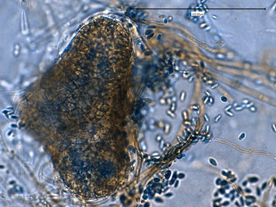
Phoma glomerata - Pycnidia seen as darkly pigmented bodies.
(LPCB, 250+10X)
Phoma glomerata - Three pycnidia showing their brown pigmentation in a LPCB preparation.
(LPCB, 400X)
Phoma glomerata - not the greatest photo example, but I like it. Pycnidium showing a massive release of conidia from within.
(LPCB, 400+10X, 12 days)
Phoma glomerata - as above. LPCB prep, however the cells are so dense that the stain hasn't penetrated throughout the preparation.
(LPCB, 400X)
Phoma glomerata - a pyriform shaped pycnidium is seen with the ostiole at center left. Interior structure of the pycnidium can be somewhat visualized through the wall. Conidiophores line the interior wall of the pycnidium where conidia are produced. Free conida can be seen throughout the photo. Pigmented septate hyphae are also seen in the background.
(LPCB, 400+10X)
Phoma glomerata - three, possibly four pycnidia. Septations are clearly visible in the hyphae at the center of the photograph.
(LPCB, 400X)
Phoma glomerata - possible small ostiole seen (arrow)
(LPCB, 400+10X, 5 days)
Phoma glomerata - Pycnidium with prominent ostiole visible, trailing conidia.
(LPCB, 400+10X, 12 days)
Phoma glomerata (LPCB, 400X)
Pathogenicity: Phoma
species are known to be pathogenic to some plants however both human and animal
infection is infrequent. Phoma glomerata has been implicated in a few documented cases of
phaeohyphomycosis. It has also been
reportedly isolated from an ulcerated human cornea.
Caution: The pycnidia produced by Phoma species should
not be confused with the perethecia suchas those of Chaetomium species or the cleistothecia of Pseudallescheria boydii.
[i] An
often flask shaped conidiomata of fungal tissue which is lined on the inside
with conidiophores. An asexual fruiting
body.
* * *
Labels:
chlamydoconidia,
Chlamydospores,
muriform,
ostiole,
Phoma,
Phoma glomerata,
pycnidia
Sunday, 14 April 2013
Trichophyton mentagrophytes Complex
Trichophyton mentagrophytes Complex (Fungus,
Dermatophyte)
Ecology: T.mentagrophytes
is recognized as having two variants.
Anthropophilic isolates prefer man to animals while the zoophilic
isolates primarily infect animals. Small
rodents appear to be the primary reservoir for the animal variety. Trichophyton
mentagrophytes is a cosmopolitan fungus (found everywhere).
Macroscopic
Morphology: T.mentagrophytes exhibits moderately rapid growth and matures
within 6 – 10 days. Sources have
previously described anthropophilic isolates having a downy, powdery or even
fluffy texture while zoophilic isolated were more granular in appearance. Colonies may vary in colour from white to cream
or yellowish. The reverse also can vary
from yellow to reddish brown to brown or ochre, depending on isolate and
medium.
Trichophyton mentagrophytes -5 days growth on SAB at 30oC
Trichophyton mentagrophytes - 14 days growth on SAB at 30oC
Microscopic
Morphology: Trichophyton mentagrophytes produces septate hyphae from which
branched conidiophores extend. Sessile (not on stalk) microconidia are produced
in rather dense, grape-like clusters on the conidiophores. The microconidia (~2µm to 4µm) are spherical
to pyriform in shape. Macroconidia (20-50µm
to 6-8µm) are cigar to club shaped and may show exhibit some distortion. Macroconidia have a smooth exterior and are
thin walled, usually have between 3 to 8 cells dividing the interior. Macroconidia may be found more readily in
younger cultures. Production of both
micro & macro conidia may vary with the isolate. Coiled or spiral hyphae may be present and in
some strains, structures described as nodular bodies or chlamydospores may be
present.
Note: All photos which follow were taken with the DMD-108 digital microscope.
T.mentagrophytes showing sessile microconidia along septate hyphae.
Note 100µm bar in upper right of this and several other photos.
(400x, LPCB)
Trichophyton mentagrophytes - branched conidiophores bearing spherical conidia in clusters seen extending from septate hyphae. (400x, LPCB)
T.mentagrophytes - another view as above. A macroconidium can be seen near the lower center of the photo (400x, LPCB)
T.metagrophytes - a closer look at the branched condiophores bearing clusters of spherical microconidia. Septations are visible in the hyphae and conidiophores.
(1000x, LPCB)
T.mentagrophytes -another look as in the previous photo.
(1000x, LPCB)
T.mentagrophytes - A 7-celled macroconidium. Macroconidia are typically described as being cigar shaped or club shaped.
(400x, LPCB)
T.mentagrophytes - another 7-celled macroconidium with dimensions (inset)
(400+10x, LPCB)
T.mentagrophytes -an macroconidium which shows slight distortion (sides are not straight). Numerous spherical microconidia in lower right.
(1000x, LPCB)
T.mentagrophytes - a solitary cigar shaped macroconidium showing seven internal cells. Walls are rather thin and the exterior is smooth.
(1000+10x, LPCB)
T.mentagrophytes - just by chance all this, and the previous macroconida all contain seven cells. I found that young cultures (~3 days) produced the most macroconidia which seemed to diminish with additional incubation. That said, the macroconidium pictured here is from a 6 day old slide culture. It should always be kept in mind that structures may appear, disappear, develop or change with length of incubation. It may be advisable to make several side cultures and harvest them at different time periods to observe development of structures.
(1000+10x, LPCB)
T.mentagrophytes - a couple of macroconidia are seen in this photo as well as clusters of microconidia. A spiral hyphal element is seen in the upper center of the photo.
(400+10x, LPCB)
T.mentagrophytes - a spiral hyphae is seen seen in the center left of this photo as it overlaps a macroconidium. Microconidia throughout the photo.
(1000x, LPCB)
T.mentagrophytes - more examples of spiral hyphae typical to T.mentagrophytes seen in this photo.
(400+10x, LPCB, 8 days incubation)
T.mentagrophytes - one last photo showing what is described as a nodular body or chlamydospore. Clusters of microconidia seen in center-left.
(1000+10x, LPCB, 10 days incubation)
Physiological
Tests: a number of classical tests can
be employed to speciate Trichophyton
species.
·
Urease test: Positive
·
BCP-Milk Solids Glucose: Alkaline reaction
·
Hair perforation test: Positive
·
Growth at 37oC: Excellent
·
Growth factor requirement*: None
*a variety of Trichophyton
tubed agars are commercially available containing specific growth supplements
(eg.inositol, thiamine, nicotinic acid, histidine). The pattern or degree of growth in each can
assist the speciation of Trichophyton.
Pathogenicity: The anthropophilic strains are usually
associated with chronic infections of glabrous skin, scalp, beard, nails and
feet.
It is currently recommended* that Trichophyton mentagrophytes be reported as ‘Trichophyton mentagrophytes complex’ which also includes the
former Trichophyton krajdenii.
*Best Practice by QMP-LS, external quality assessment
agency.
* * *
Saturday, 13 April 2013
Trichothecium roseum
Trichothecium roseum (Fungus)
Ecology: Trichothecium species are cosmopolitan
fungi (found just about everywhere) and is a common saprobe (growing on
decaying vegetation). In particular, it
has been isolated from withering fleshy fruits such as peaches, plums and
nectarines. Trichothecium is responsible for ‘pink rot’ of apples.
Macroscopic
Morphology: Trichothecium is a rapidly growing fungus which is fully mature in
3 to 4 days. Colonies are described as
being flat, suede-like to powdery, which start off white but quickly develop a
light pink to peach colour which may reach a darker salmon colour on continued
incubation. The reverse is a rather non-descript
light or pale colour. Pictured below are
colonies grown at 30oC on Sabouraud Dextrose Media (SAB or SDA). Trichothecium
fails to grow at 37oC making it an unlikely candidate for human
pathogenicity.
Trichothecium roseum on SAB agar, 72 hrs, 30oC (Nikon)
It was rather difficult to capture (and correct for) the exact colour hue under harsh labor fluorescent laboratory lighting within the biological laminar flow hood. The colour descriptions range from a light pink to peachy to salmon or even orange on extended incubation.
Trichothecium roseum on SAB after 14 days incubation at 30oC (Nikon)
Microscopic
Morphology: Hyphae produced by Trichothecium are septate and hyaline
(clear, not pigmented). The long, thin and
erect conidiophores are indistinguishable from the vegetative hyphae and may
exhibit septation near their base of attachment. Trichothecium
roseum produces rather thin walled, two-celled conidia (16-20µm X 8-12µm)
which are pyriform or clavate in shape.
Basipetal growth has the newest cell developing below the previous one (The youngest cells are at the base while
the oldest are at the apex.) This
growth produces a sympoidal pattern seen as zigzag or alternating conidia extending
from the conidiophore on opposite sides.
Free conidia have a truncated basal scar usually obliquely offset,
indicating their former point of attachment.
Note: All photos which appear below were taken with the Leica DMD-108 digital microscope.
Trichothecium roseum growing from the surface edge of agar (bottom of photo). Fine hyphae and conidiophores bearing conidia are seen (7 days 250X LPCB)
Trichothecium roseum -conidiophores are seen extending along the lengths of hyphae. Conidia are seen clumped at the apex of the hyphae. Branching of conidiophores is rare if it occurs at all.
(250x. LPCB)
Trichothecium roseum - conidiophores with early production of two-celled conidia are seen extending from a hyphal element just out of focus below. A number of two-celled conidia are seen free of the conidiophore. A truncated basal scare can be seen which may may be somewhat offset from center (arrow), due to the sympodial pattern of growth.
(400X, LPCB)
Trichothecium roseum - again, conidiophores are seen extending from the hyphae (slightly out of focus at top). Cells appear 'clustered' around the apex of the conidiophore as the growth extends. Note 100µm bar at top right of this and previous photo.
(LPCB, 400X)
Trichothecium roseum - conidiophores extending from hyphae with pyrimidal or clavate shaped (pear shaped) conidia extending to the apex. Again, 100µm bar appears on this and various other photos for scale. (400X, LPCB)
Trichothecium roseum - somewhat thick walled, two-celled clavate conidia are seen at the apex of the conidiophore extending upwards into the focal plane of the camera. The conidium at the top of the group appears contorted (twisted) at the bottom where it is attached to the conidiophore. When released, a truncated basal scar will be present at this attachment point and it will be somewhat offset from the centerline of the conidium. (1000X, LPCB)
Trichothecium roseum - once again the clavate shaped conidia are seen attached along the conidiophore. Here you can see the sympodial growth pattern which produces conidia in an alternating or zigzag pattern along the conidiophore. The youngest cells are at the bottom with the most mature at the apex (top) of the cluster. (1000X, LPCB)
Trichothecium roseum - free conidia showing the pyramidal, clavate (club shaped) or perhaps pear shape characteristic of this fungus. The cell wall is rather thin to moderately thickened and the central division is visible in most cells above. Again, the basal scar appears at the previous point of attachment and may be somewhat offset from the center line of the conidium.
(1000X, LPCB)
Trichothecium roseum - clavate two-celled conidia seen attached to conidiophore
(1000+10X, LPCB)
Trichothecium roseum - one more photo just for the heck of it. Septate hyphae can be seen. Sympodial attachment of cells can be seen with the two cells near the center of the photo.
(400X, 14 days, LPCB)
Trichothecium roseum -Computer wallpaper (1024X768)
Pathogenicity: Trichothecium species are generally clinical
laboratory contaminants. No human or
animal infections have been reported.
Differentiation: Trichothecium
roseum may initially be confused with Microsporum
nanum as this fungus produces a similar light pink to buff coloration. M.nanum,
however, exhibits a reddish-brown pigment on reverse in contrast to the pale
reverse of T.roseum. The conidia produced by M.nanum are also two celled however they are sessile (attached directly
to undifferentiated conidiophores) or on short stalks. Finally, Microsporum nanum has the ability to
perforate hair cells and is not inhibited by the cycloheximide in Mycosel agar.
* * *
Subscribe to:
Comments (Atom)
+5+days.jpg)
+14+days.jpg)
.jpg)


.jpg)
.jpg)
.jpg)


.jpg)
.jpg)
+14d.jpg)


.jpg)

.jpg)


